
Page 136 - TNFlipTest
P. 136
D2 Dermatology Acronyms
β-hCG β-human chorionic gonadotropin
AAFP American Academy of Family Physicians AGEP acute generalized
exanthematous pustulosis AD atopic dermatitis
AK actinic keratosis
ASO anti-streptolysin O
BCC basal cell carcinoma
BSA body surface area
BUN blood urea nitrogen
CBC complete blood count
CMV cytomegalovirus
CNS central nervous system
Cr creatinine
CXR chest x-ray
DIHS drug induced hypersensitivity syndrome DLE discoid lupus erythematosus
DM diabetes mellitus
DRESS drug reaction with eosinophilia and
Acronyms Toronto Notes 2019
Fe iron aureus SLE systemic lupus erythematosus FTA-ABS fluorescent treponemal MTP metatarsal phalangeal SPF sun protection factor
antibody-absorption NB-UVB narrow band ultraviolet wavelength B SSRI selective serotonin reuptake inhibitor GAS group A β-hemolytic Streptococcus Nd:YAG neodymium-doped yttrium aluminum SSSS staphylococcal scalded skin
GVHD graft-versus-host disease garnet syndrome
HHV human herpes virus NMN nevomelanocytic nevus STI sexually transmitted infection
HPA hypothalamic-pituitary-adrenal NMSC nonmelanoma skin cancers TB tuberculosis
HPV human papillomavirus NSAID nonsteroidal anti-inflammatory drug TEN toxic epidermal necrolysis
HRT hormone replacement therapy OCP oral contraceptive pill TMP/SMX trimethoprim-sulfamethoxazole
HSV herpes simplex virus OTC over-the-counter TSH thyroid stimulating hormone
HZV herpes zoster virus PABA para-aminobenzoic acid UC ulcerative colitis
IFN interferon PASI psoriasis area and severity index URTI upper respiratory tract infection
IVIg intravenous immunoglobulin PPD purified protein derivative UV ultraviolet
LFT liver function test PUVA psoralens and UVA UVA ultraviolet A
MAOI monoamine oxidase inhibitor RA rheumatoid arthritis UVB ultraviolet B
MM malignant melanoma SCC squamous cell carcinoma UVC ultraviolet C
MMR measles/mumps/rubella SHBG sex hormone-binding globulin VDRL venereal disease research laboratory MRSA methicillin-resistant Staphylococcus SJS Stevens-Johnson syndrome VZV varicella zoster virus
systemic symptoms DVT deep vein thrombosis EM erythema multiforme
Layers of the Epidermis “Californians Like Getting Sun Burns”
OR
“Canadians Like Good Sushi Boxes”
Introduction to Skin
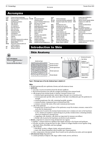
Skin Anatomy A
Epidermal layer
Arrector pili muscle Hair follicle
Epidermis Papillary layer
Dermis
Reticular layer
Subcutaneous adipose tissue
Sweat gland
◆ stratum basale (germinativum): mitotic figures that give rise to keratinocytes
◆ stratum spinosum (prickle cells): junctions in this layer (tonofilaments) give the epidermis its
strength
◆ stratum granulosum: flat cells containing basophilic granules
◆ stratum lucidum: transparent layers of packed dead cells
◆ stratum corneum: flat scales of the water-resistant protein keratin
■ cells of the epidermis
◆ keratinocytes: located in all layers of the epidermis, except the stratum corneum; connected to
each other by desmosomes
◆ melanocytes: located in the stratum basale; keratinocyte to melanocyte ratio in the basal layer
is 10:1; melanocyte number is equal among races; produce melanosomes containing melanin,
which are transferred to keratinocytes
◆ Langerhans cells: dendritic cells which are important for immune surveillance ◆ Merkel cells: located in the basal layer; involved in touch sensation
• dermis:comprisedofconnectivetissuedividedintotworegions
■ papillary: contains numerous capillaries that supply nutrients to the dermis and epidermis
■ reticular: provides a strong structure for skin; consists of collagen bundles woven together along
with elastic fibres, fibroblasts, and macrophages
■ cells of dermis
◆ fibroblasts: produce collagen, elastin, and ground substance
◆ mast cells: release histamines which mediate type I hypersensitivity
■ other components of dermis include: blood vessels, nerves, pilosebaceous units, and sweat glands
• subcutaneoustissue(hypodermis)
■ consists primarily of adipose cells, larger calibre vessels, nerves, and fascia
B
Stratum corneum Stratum lucidum Stratum granulosum
Stratum spinosum Stratum basale
Figure 1. Histologic layers of the skin. Epidermal layer is detailed in A
Skin
• dividedanatomicallyintoepidermis,dermis,andsubcutaneoustissue • epidermis
■ avascular: receives its nutrition from the dermal capillaries
■ derived from keratinocytes with the youngest presenting at the stratum basale ■ cellsprogressfromstratumbasaletostratumcorneuminabout4wk
© Ken Vanderstoep 2002
