
Page 191 - TNFlipTest
P. 191
Toronto Notes 2019
Traumatology
Emergency Medicine ER11
3. AP view
◆ alignment of spinous processes in the midline ◆ spacing of spinous processes should be equal ◆ check vertebral bodies and facet dislocations
Table 6. Interpretation of Lateral View: The ABCS
A Adequacy and Alignment
Must see C1 to C7-T1 junction; if not, downward traction of shoulders, swimmer’s view, bilateral supine obliques, or CT scan needed
Lines of contour in children <8 yr of age, can see physiologic subluxation of C2 on C3, and C3 on C4, but the spino-laminal line is maintained
Fanning of spinous processes suggests posterior ligamentous disruption
Widening of facet joints
Check atlanto-occipital joint
Line extending inferiorly from clivus should transect odontoid
Atlanto-axial articulation, widening of predental space (normal: <3 mm in adults, <5 mm in children) indicates injury of C1 or C2
B Bones
Height, width, and shape of each vertebral body
Pedicles, facets, and laminae should appear as one – doubling suggests rotation
C Cartilage
Intervertebral disc spaces – wedging anteriorly or posteriorly suggests vertebral compression
S Soft Tissues
Widening of retropharyngeal (normal: <7 mm at C1-4, may be wide in children <2 yr on expiration) or retrotracheal spaces (normal: <22 mm at C6-T1, <14 mm in children <5 yr)
Sequelae of C-Spine Fractures
• seeNeurosurgery,NS34 • acutephaseofSCI
■ spinal shock: absence of all voluntary and reflex activity below level of injury
◆ decreased reflexes, no sensation, flaccid paralysis below level of injury, lasting days to months
■ neurogenic shock: loss of vasomotor tone, SNS tone
◆ watch for: hypotension (lacking SNS), bradycardia (unopposed PNS), poikilothermia (lacking
SNS so no shunting of blood from extremities to core)
◆ occurs within 30 min of SCI at level T6 or above, lasting up to 6 wk
◆ provide airway support, fluids, atropine (for bradycardia), vasopressors for BP support
• chronicphaseofSCI
■ autonomic dysreflexia: in patients with an SCI at level T6 or above
◆ signs and symptoms: pounding headache, nasal congestion, feeling of apprehension or anxiety, visual changes, dangerously increased sBP and dBP
◆ common triggers
– GU causes: bladder distention, urinary tract infection, and kidney stones – GI causes: fecal impaction or bowel distension
◆ treatment: monitoring and controlling BP, prior to addressing causative issue
Chest Trauma
• twotypes:thosefoundandmanagedin1°surveyandthosefoundandmanagedin2°survey
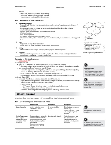
2 1 2 3
1. Dens of C2
2. C1 lateral mass 3. C2
To clear the x-ray ensure that:
A. The dens is centred between the
lateral masses of C1
B. C1 and C2 are aligned laterally
C. The lateral masses of C1 are
symmetrical in size
Figure 5. C-spine x-ray; odontoid view
Supine Oblique Views
• Rarely used
• Better visualization of posterior element
fractures (lamina, pedicle, facet joint)
• Good to assess patency of neural foramina • Can be used to visualize the C7-T1 junction
20% of C-spine fractures are accompanied by other spinal fractures, so ensure thoracic and lumbar spine x-rays are normal before proceeding to OR
Table 7. Life-Threatening Chest Injuries Found in 1o Survey
Airway Obstruction
Tension Pneumothorax
Clinical diagnosis One-way valve causing accumulation of air in pleural space
Physical Exam
Anxiety, stridor, hoarseness, altered mental status Apnea, cyanosis
Respiratory distress, tachycardia, distended neck veins, cyanosis, asymmetry of chest wall motion Tracheal deviation away from pneumothorax
Percussion hyperresonance Unilateral absence of breath sounds
Investigations
Do not wait for ABG to intubate
Non-radiographic diagnosis
Management
Definitive airway management Intubate early
Remove foreign body if visible with laryngoscope prior to intubation
Needle thoracostomy – large bore needle, 2nd ICS mid clavicular line, followed by chest tube in 5th ICS, anterior axillary line
Trauma to the chest accounts for 50% of trauma deaths
80% of all chest injuries can be managed non-surgically with simple measures such as intubation, chest tubes, and pain control
© Eddy Xuan
