
Page 192 - TNFlipTest
P. 192
ER12 Emergency Medicine
Traumatology
Toronto Notes 2019
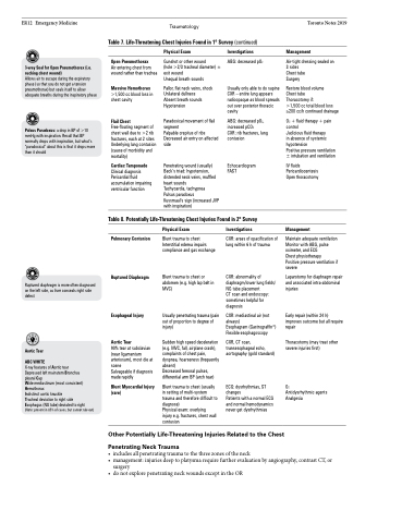
Table 7. Life-Threatening Chest Injuries Found in 1o Survey (continued)
3-way Seal for Open Pneumothorax (i.e. sucking chest wound)
Allows air to escape during the expiratory phase (so that you do not get a tension pneumothorax) but seals itself to allow adequate breaths during the inspiratory phase
Pulsus Paradoxus: a drop in BP of >10 mmHg with inspiration. Recall that BP normally drops with inspiration, but what’s “paradoxical” about this is that it drops more than it should
Open Pneumothorax
Air entering chest from wound rather than trachea
Massive Hemothorax
>1,500 cc blood loss in chest cavity
Flail Chest
Free-floating segment of chest wall due to >2 rib fractures, each at 2 sites Underlying lung contusion (cause of morbidity and mortality)
Cardiac Tamponade
Clinical diagnosis Pericardial fluid accumulation impairing ventricular function
Physical Exam
Gunshot or other wound
(hole >2/3 tracheal diameter) ± exit wound
Unequal breath sounds
Pallor, flat neck veins, shock Unilateral dullness
Absent breath sounds Hypotension
Paradoxical movement of flail segment
Palpable crepitus of ribs Decreased air entry on affected side
Penetrating wound (usually) Beck’s triad: hypotension, distended neck veins, muffled heart sounds
Tachycardia, tachypnea
Pulsus paradoxus
Kussmaul’s sign (increased JVP with inspiration)
Investigations
ABG: decreased pO2
Usually only able to do supine CXR – entire lung appears radioopaque as blood spreads out over posterior thoracic cavity
ABG: decreased pO2, increased pCO2
CXR: rib fractures, lung contusion
Echocardiogram FAST
Management
Air-tight dressing sealed on 3 sides
Chest tube
Surgery
Restore blood volume
Chest tube
Thoracotomy if:
>1,500 cc total blood loss ≥200 cc/h continued drainage
O2 + fluid therapy + pain control
Judicious fluid therapy
in absence of systemic hypotension
Positive pressure ventilation ± intubation and ventilation
IV fluids Pericardiocentesis Open thoracotomy
Management
Maintain adequate ventilation Monitor with ABG, pulse oximeter, and ECG
Chest physiotherapy
Positive pressure ventilation if severe
Laparotomy for diaphragm repair and associated intra-abdominal injuries
Early repair (within 24 h) improves outcome but all require repair
Thoracotomy (may treat other severe injuries first)
O2
Antidysrhythmic agents Analgesia
Table 8. Potentially Life-Threatening Chest Injuries Found in 2o Survey
Ruptured diaphragm is more often diagnosed on the left side, as liver conceals right side defect
Aortic Tear
ABC WHITE
X-ray features of Aortic tear Depressed left mainstem Bronchus pleural Cap
Wide mediastinum (most consistent) Hemothorax
Indistinct aortic knuckle
Tracheal deviation to right side
Esophagus (NG tube) deviated to right (Note: present in 85% of cases, but cannot rule out)
Pulmonary Contusion
Ruptured Diaphragm
Esophageal Injury
Aortic Tear
90% tear at subclavian (near ligamentum arteriosum), most die at scene
Salvageable if diagnosis made rapidly
Blunt Myocardial Injury (rare)
Physical Exam
Blunt trauma to chest Interstitial edema impairs compliance and gas exchange
Blunt trauma to chest or abdomen (e.g. high lap belt in MVC)
Usually penetrating trauma (pain out of proportion to degree of injury)
Sudden high speed deceleration (e.g. MVC, fall, airplane crash), complaints of chest pain, dyspnea, hoarseness (frequently absent)
Decreased femoral pulses, differential arm BP (arch tear)
Blunt trauma to chest (usually in setting of multi-system trauma and therefore difficult to diagnose)
Physical exam: overlying injury e.g. fractures, chest wall contusion
Investigations
CXR: areas of opacification of lung within 6 h of trauma
CXR: abnormality of diaphragm/lower lung fields/ NG tube placement
CT scan and endoscopy: sometimes helpful for diagnosis
CXR: mediastinal air (not always)
Esophagram (Gastrograffin®) Flexible esophagoscopy
CXR, CT scan, transesophageal echo, aortography (gold standard)
ECG: dysrhythmias, ST changes
Patients with a normal ECG and normal hemodynamics never get dysrhythmias
Other Potentially Life-Threatening Injuries Related to the Chest
Penetrating Neck Trauma
• includesallpenetratingtraumatothethreezonesoftheneck
• management:injuriesdeeptoplatysmarequirefurtherevaluationbyangiography,contrastCT,or
surgery
• donotexplorepenetratingneckwoundsexceptintheOR
