
Page 525 - TNFlipTest
P. 525
Toronto Notes 2019 Gynecological Oncology
Screening
• noknownbenefitformassscreening
• routinepelvicultrasoundshouldnotbeusedasscreeningtest(highfalsepositives)
Investigations
• endometrialsampling
■ office endometrial biopsy ■ D&C±hysteroscopy
• ±pelvicultrasound(inwomenwhereadequateendometrialsamplingnotfeasiblewithoutinvasive methods)
■ not acceptable as alternative to pelvic exam or endometrial sampling to rule out cancer
Gynecology GY39
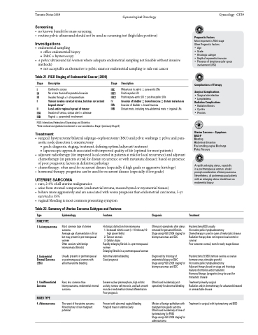
Table 21. FIGO Staging of Endometrial Cancer (2009)
Stage Description
I Confined to corpus
IA No or less than half myometrial invasion
IB Invades through ≥1⁄2 of myometrium
II Tumour invades cervical stroma, but does not extend
beyond uterus*
III Local and/or regional spread of tumour
IIIA Invasion of serosa, corpus uteri ± adnexae
IIIB Vaginal ± parametrial involvement
Stage Description
IIIC Metastasis to pelvic ± para-aortic LNs
IIIC1 Positive pelvic LN
IIIC2 Positive para-aortic LN ± positive pelvic LNs
IV Invasion of bladder ± bowel mucosa ± distant metastases
IVA Invasion of bladder ± bowel mucosa
IVB Distant mets, including intra-abdominal mets ± inguinal LNs
Prognostic Factors
Most important is FIGO stage Other Prognostic Factors:
• Age
• Grade
• Histologic subtype
• Depth of myometrial invasion
• Presence of lymphovascular space
involvement (LVSI)
Complications of Therapy Surgical Complications
• Surgical site infection • Lymphedema Radiation Complications • Radiation fibrosis
• Cystitis • Proctois
Uterine Sarcoma – Symptoms BAD-P
Bleeding
Abdominal distention
Foul smelling vaginal Discharge Pelvic Pressure
A rapidly enlarging uterus, especially
in a postmenopausal woman, should prompt consideration of leiomyosarcoma. Nevertheless, all postmenopausal patients with an enlarging uterus should have an endometrial biopsy
Treatment
Hysterectomy/BSO usually
No routine pelvic lymphadenectomy
Chemotherapy is used in cases of metastatic disease Radiation therapy does not improve local control or survival
Poor outcomes overall, even for early stage disease
Hysterectomy & BSO (remove ovaries as ovarian hormones may stimulate growth)
No routine pelvic lymphadenectomy
Adjuvant therapy based on stage and histologic features (hormones and/or radiation)
Hormonal therapy (progestins) may be used for metastatic disease
Treatment primarily surgical
Radiation and/or chemotherapy for advanced diseased or unresectable disease
Treatment is surgical with hysterectomy and BSO
FIGO: International Federation of Gynecology and Obstetrics
*Note: endocervical glandular involvement is now considered as Stage I (previously Stage II)
Treatment
• surgical:hysterectomy/bilateralsalpingo-oophorectomy(BSO)andpelvicwashings±pelvicandpara- aortic node dissection ± omentectomy
■ goals: diagnosis, staging, treatment, defining optimal adjuvant treatment
■ laparoscopic approach associated with improved quality of life (optimal for most patients)
• adjuvantradiotherapy(forimprovedlocalcontrolinpatientsatriskforlocalrecurrence)andadjuvant chemotherapy (in patients at risk for distant recurrence or with metastatic disease): based on presence
of poor prognostic factors in definitive pathology
• chemotherapy:oftenusedforrecurrentdisease(especiallyifhighgradeoraggressivehistology)
• hormonaltherapy:progestinscanbeusedforrecurrentdisease(especiallyiflowgrade)
UTERINE SARCOMA
• rare;2-6%ofalluterinemalignancies
• arisefromstromalcomponents(endometrialstroma,mesenchymalormyometrialtissues)
• behavemoreaggressivelyandareassociatedwithworseprognosisthanendometrialcarcinoma;5-yr
survival is 35%
• vaginalbleedingismostcommonpresentingsymptom
Table 22. Summary of Uterine Sarcoma Subtypes and Features
Type
1. Leiomyosarcoma
2. Endometrial Stromal Sarcoma (ESS)
3. Undifferentiated Sarcoma
4. Adenosarcoma
Epidemiology
Most common type of uterine sarcoma
Average age of presentation is 55 yr but may present in pre-menopausal women
Often coexists with benign leiomyomata (fibroids)
Usually presents in perimenopausal or postmenopausal women with abnormal uterine bleeding
Rare; less common than leiomyosarcoma, endometrial stromal sarcoma
The rarest of the uterine sarcoma Mixed tumour of low malignant potential
Features
Histologic distinction from leiomyoma
1. Increased mitotic count (>10 mitoses/10
high power fields) 2. Tumour necrosis 3. Cellular atypia
Rapidly enlarging fibroids in a pre-menopausal woman
Enlarging fibroids in a postmenopausal woman
Abnormal uterine bleeding Good prognosis
Severe nuclear pleomorphism, high mitotic activity, tumour cell necrosis, and lack smooth muscle or endometrial stromal differentiation Poor prognosis
Present with abnormal vaginal bleeding Polypoid mass in uterine cavity
Diagnosis
Often post-operatively after uterus removed for presumed fibroids Stage using FIGO 2009 staging for leiomyosarcomas and ECC
Diagnosed by histology of endometrial biopsy or D&C
Stage using FIGO 2009 staging for leiomyosarcomas and ECC
Often found incidentally post- operatively for abnormal bleeding
Mixture of benign epithelium with malignant low-grade sarcoma Often found incidentally at time of hysterectomy for PMB
Stage using FIGO 2009 staging for adenosarcoma
PURE TYPE
MIXED TYPE
