
Page 528 - TNFlipTest
P. 528
GY42 Gynecology
Gynecological Oncology
Toronto Notes 2019
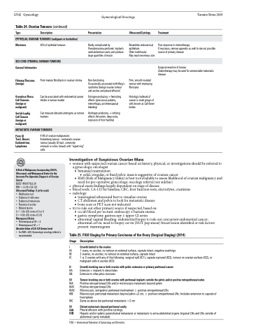
Table 24. Ovarian Tumours (continued)
Type Description
EPITHELIAL OVARIAN TUMOURS (malignant or borderline)
Presentation
Rarely complicated by Pseudomyxoma peritoneii: implants seed abdominal cavity and produce large quantities of mucin
Non-functioning
Occasionally associated with Meig’s syndrome (benign ovarian tumour and ascites and pleural effusion)
Estrogen-producing → feminizing effects (precocious puberty, menorrhagia, postmenopausal bleeding)
Androgen-producing → virilizing effects (hirsutism, deep voice, recession of front hairline)
Ultrasound/Cytology
Resembles endocervical epithelium
Often multilocular
May reach enormous size
Firm, smooth rounded tumour with interlacing fibrocytes
Histologic hallmark of cancer is small groups of cells known as Call-Exner bodies
Treatment
Poor response to chemotherapy
If mucinous, remove appendix as well to rule out possible source of primary disease
Surgical resection of tumour
Chemotherapy may be used for unresectable metastatic disease
Mucinous
General Information
Fibroma/Thecoma (benign)
Granulosa-Theca Cell Tumours (benign or malignant)
Sertoli-Leydig Cell Tumour (benign or malignant)
From GI Tract, Breast, Endometrium, Lymphoma
20% of epithelial tumours
From mature fibroblasts in ovarian stroma
Can be associated with endometrial cancer Inhibin is tumour marker
Can measure elevated androgens as tumour markers
4-8% of ovarian malignancies
Krukenberg tumour – metastatic ovarian tumour (usually GI tract, commonly stomach or colon, breast) with “signet-ring” cells
SEX CORD STROMAL OVARIAN TUMOURS
METASTATIC OVARIAN TUMOURS
Investigation of Suspicious Ovarian Mass
• womenwithsuspectedovariancancerbasedonhistory,physical,orinvestigationsshouldbereferredto a gynecologic oncologist
A Risk of Malignancy Incorporating CA125, Ultrasound, and Menopausal Status for the Accurate Pre-Operative Diagnosis of Ovarian Cancer
BJOG 1990;97:922-29
RMI=UxMxCA-125
Ultrasound Findings (1 pt for each)
• Multilocularcyst
• Evidenceofsolidareas
• Evidenceofmetastases
• Presenceofascites
• Bilaterallesions U=1(forU/Sscoresof0or1)
U = 4 (for U/S scores of 2-5)
Menopausal Status
• Postmenopausal:M=4
• Premenopausal:M=1
Absolute Value of CA-125 Serum Level
• ForRMI>200:Gynecologiconcologyreferralis
recommended
■ bimanual examination
◆ solid, irregular, or fixed pelvic mass is suggestive of ovarian cancer
■ RMI (Risk of Malignancy Index) is best tool available to assess likelihood of ovarian malignancy and need for pre-operative gynecologic oncology referral (see sidebar)
• • •
•
Table 25. FIGO Staging for Primary Carcinoma of the Ovary (Surgical Staging) (2014) Stage Description
I Growth limited to the ovaries
IA 1 ovary, no ascites, no tumour on external surface, capsule intact, negative washings
IB 2 ovaries, no ascites, no tumour on external surface, capsule intact
IC 1 or 2 ovaries with any of the following: surgical spill (IC1), capsule ruptured (IC2), tumour on ovarian surface (IC2), or
malignant cells in ascites (IC3)
II Growth involving one or both ovaries with pelvic extension or primary peritoneal cancer
IIA Extension ± implants to uterus/tubes
IIB Extension to other pelvic structures
III Tumour involving one or both ovaries with peritoneal implants outside the pelvis and/or positive retroperitoneal nodes
IIIA Positive retroperitoneal LNs and/or microscopic metastasis beyond pelvis
IIIA1 Positive retroperitoneal LNs
IIIA2 Microscopic, extrapelvic peritoneal involvement ± positive retroperitoneal LNs
IIIB Macroscopic peritoneal metastasis beyond pelvis ≤2 cm, ± positive retroperitoneal LNs. Includes extension to capsule of
liver/spleen
IIIC Same as above but peritoneal metastasis >2 cm
IV Distant metastasis beyond peritoneal cavity
IVA Pleural effusion with positive cytology
IVB Hepatic and/or splenic parenchymal metastasis or metastasis to extra-abdominal organs (inguinal LNs and LNs outside of
abdominal cavity included)
FIGO = International Federation of Gynecology and Obstetrics
physicalexamfindingslargelydependentonstageofdisease bloodwork:CA-125forbaseline,CBC,liverfunctiontests,electrolytes,creatinine radiology
■ transvaginal ultrasound best to visualize ovaries
■ CT abdomen and pelvis to look for metastatic disease ■ bone scan or PET scan not indicated
try to rule out other primary source if suspected, based on:
■ occult blood per rectum: endoscopy ± barium enema
■ gastric symptoms: gastroscopy ± upper GI series
■ abnormal vaginal bleeding: endometrial biopsy to rule out concurrent endometrial cancer;
abnormal cervix: need to biopsy cervix (NOT pap smear) breast lesion identified or risk factors present: mammogram
