Page 590 - TNFlipTest
P. 590
 H50 Hematology
Malignant Clonal Proliferations of Mature B-Cells
Toronto Notes 2019
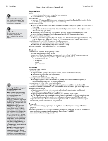
  Routine urinalysis will not detect light chains as dipstick detects albumin. Need sulfosalicylic acid or 24 h urine protein for immunofixation or electrophoresis
Light Chain Disease
15% of MM produce only light chains Renal failure is a major problem
Kappa > lambda light chain has better prognosis
Investigations
• CBC
■ normocytic anemia, thrombocytopenia, l and eukopenia ■ rouleaux formation on peripheral film
• biochemistry
■ increased Ca2+, increased ESR, decreased anion gap, increased Cr, albumin, β2-microglobulin (as
part of staging), and proteinuria (24 h urine collection) • monoclonalproteins
■ serum protein electrophoresis (SPEP): demonstrates monoclonal protein spike in serum in 80% (i.e. M protein)
■ urine protein electrophoresis (UPEP): demonstrates light chains in urine = Bence-Jones protein (15% secrete only light chains)
■ immunofixation: demonstrates M protein and identifies Ig type; also identifies light chains
■ serum free light chain quantification: kappa and lambda light chains, calculated ratio • bonemarrowaspirateandbiopsy
■ often focal abnormality, greater than 10% plasma cells, abnormal morphology, clonal plasma cells; send for fluorescence in situ hybridization (FISH) or cytogenetics (prognostic implications)
• skeletalseries(X-rays),MRIifsymptomsofcordcompression
■ presence of lytic lesions and areas at risk of pathologic fracture ■ bone scans are not useful since they detect osteoblast activity
• β2-microglobulin,LDH,andCRParepoorprognosticators
Diagnosis
• InternationalMyelomaWorkingGroupCriteria
1. serum or urinary monoclonal protein
2. presence of clonal plasma cells in bone marrow (>60% without “CRAB”) or a plasmacytoma 3. presence of end-organ damage related to plasma cell dyscrasia, such as:
◆ increased serum Ca2+ ◆ renal failure
◆ anemia
◆ lytic bone lesions
Treatment
• treatmentisnon-curative
• treatmentgoals
■ improvement in quality of life (improve anemia, reverse renal failure, bony pain) ■ prevention of progression and complications
■ increase overall survival
• autologousstemcelltransplantif<65yrold
■ usually preceded by 4-6 mo of cytoreductive therapy: steroid based with novel agents (i.e.
immunomodulatory drugs or proteasome inhibitors)
• chemotherapyif>65yroldortransplant-ineligible
■ melphalan, prednisone, cyclophosphamide and proteasome inhibitor (i.e. bortezomib)
• dexamethasoneandbortezomibifARF;bortezomib±dexamethasoneinlightchainamyloidosis
• supportivemanagement
■ bisphosphonates for those with osteopenia or lytic bone lesions (requires renal dosing)
■ local XRT for bone pain, spinal cord compression
■ kyphoplasty for vertebral fractures to improve pain relief and regain height
■ treat complications: hydration for hypercalcemia and renal failure, bisphosphonates for severe
hypercalcemia, prophylactic antibiotics, erythropoietin for anemia, and DVT prophylaxis
• allpatientswillrelapse;choiceofretreatmentregimendependsondurationofremission,organ
involvement, patient’s comorbidities, and preferences
Prognosis
• ISS-InternationalStagingSystem(β2-microglobulinandalbumin)usedtostageandestimate prognosis
• revisedISSforriskstratification:combinationoforiginalISS,cytogeneticprofile(i.e.p53mutation associated with poor survival and resistance to chemotherapy), and LDH
• median survival based on stage, usually 3-7 yr
            Serum Free Light Chain Ratio is an Independent Risk Factor for Progression in MGUS
Blood 2005;106:812-817
Purpose: To determine whether the presence of monoclonal free kappa or lambda immunoglobulin light chains in MGUS increases the risk of progression to malignancy.
Methods: Retrospective study with median follow-up of 15 yr. Baseline serum samples obtained from 1,383 MGUS patients seen at the Mayo clinic between 1960-1994. 1,148 baseline samples were obtained within 30 d of diagnosis.
Results: Malignant progression had occurred
in 87 (7.6%) patients. In 379 (33%) patients, an abnormal serum free light chain (FLC) ratio was detected. There was a significantly higher risk of progression in patients with an abnormal FLC ratio relative to patients with a normal ratio (hazard
ratio, 3.5; 95% Cl 2.3-5.5; p<0.001). This finding was independent of the size and type of the serum monoclonal (M) protein. In high-risk MGUS patients (abnormal serum FLC ratio, non-IgG MGUS, high serum M protein level [≥1.5 gm/dL]), the risk of progression at 20 yr was 58% compared to 37%
in high-intermediate-risk MGUS (two risk factors), 21% low-intermediate risk (with one risk factor) and 5% low-risk (no risk factors).
Conclusions: The presence of an abnormal FLC ratio is a clinically and statistically significant predictor of progression in MGUS. The low-risk subset of patients with MGUS accounts for 40% of all MGUS patients and have a small lifetime risk of progression, thus less follow-up can be justified.


