
Page 670 - TNFlipTest
P. 670
MI2 Medical Imaging
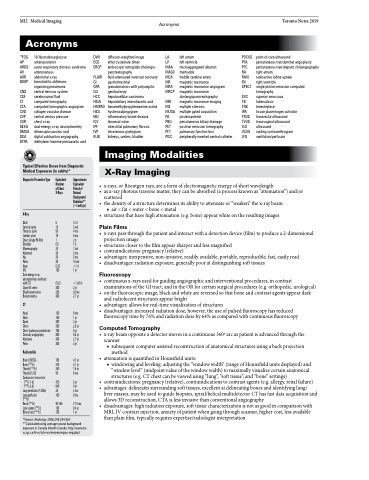
Acronyms
Toronto Notes 2019
Acronyms
18FDG 18-fluorodeoxyglucose DWI AP anteroposterior ECD ARDS acute respiratory distress syndrome ERCP AV arteriovenous
AXR abdominal x-ray FLAIR BOOP bronchiolitis obliterans GI
organizing pneumonia GPA CNS central nervous system GU CSF cerebrospinal fluid HCC CT computed tomography HIDA CTA computed tomographic angiogram HMPAO CVD collagen vascular disease HSG CVP central venous pressure IBD CXR chest x-ray ICV DEXA dual-energy x-ray absorptiometry IPF DMSA dimercaptosuccinic acid IVP DSA digital subtraction angiography KUB DTPA diethylene triamine pentaacetic acid
diffusion-weighted image
ethyl cysteinate dimer
endoscopic retrograde cholangio- pancreatography
fluid-attenuated inversion recovery gastrointestinal
granulomatosis with polyangiitis genitourinary
hepatocellular carcinoma hepatobiliary iminodiacetic acid hexamethylpropyleneamine oxime hysterosalpingogram inflammatory bowel disease ileocecal valve
interstitial pulmonary fibrosis intravenous pyelogram kidneys, ureters, bladder
LA left atrium
LV left ventricle
MAA microaggregated albumin MAG3 mertiatide
MCA middle cerebral artery
MR magnetic resonance
MRA magnetic resonance angiogram MRCP magnetic resonance
cholangiopancreatography MRI magnetic resonance imaging
MS multiple sclerosis
MUGA multiple gated acquisition
PA posteroanterior
PBD percutaneous biliary drainage
PET positron emission tomography
PFT pulmonary function test
PICC peripherally-inserted central catheter
POCUS point-of-care ultrasound
PTA percutaneous transluminal angioplasty
PTC percutaneous transhepatic cholangiography RA right atrium
RAIU radioactive iodine uptake
RV right ventricle
SPECT single photon emission computed
tomography
SVC superior vena cava
TB tuberculosis
TNK tenecteplase
tPA tissue plasminogen activator TRUS transrectal ultrasound
TVUS transvaginal ultrasound
U/S ultrasound
VCUG voiding cystourethrogram V/Q ventilation/perfusion
Typical Effective Doses from Diagnostic Medical Exposures (in adults)*
Imaging Modalities
X-Ray Imaging
Diagnostic Procedure Type
X-Ray
Skull
Cervical spine
Thoracic spine
Lumbar spine
Chest (single PA film) Shoulder Mammography Abdomen
Hip
Pelvis
Knee
IVU
Dual-energy x-ray absorptiometry (without/ with CT)
Upper GI series
Small bowel series Bariumenema
CT
Head
Neck
Spine
Chest
Chest (pulmonary embolism) Coronary angiography Abdomen
Pelvis
Radionuclide
Brain (18FDG)
Bone (99mTc)
Thyroid (99mTc)
Thyroid (123I)
Cardiac rest-stress test (99mTc 1-d)
(99mTc 2-d)
Lung ventilation (133Xe) Lung perfusion
(99mTc)
Renal (99mTc) Liver-spleen (99mTc) Biliary tract (99mTc)
Equivalent Number of Chest X-Rays
5 10 50 75 1 0.5 20 35 35 30 0.25 150
0.5/2 300 250 400
100 150 300 350 750 800 400 300
705 315 240 95
470 640 25 100
90-165 105 155
Approximate Equivalent Period of Natural Background Radiation** (~3 mSv/yr)
12 d 3 wk 4 mo 6 mo 2d 1d
7 wk 3 mo 3 mo 10 wk <1 d 1 yr
<1 d/4 d 2 yr
20 mo 2.7 yr
8 mo 1 yr 2 yr 2.3 yr 5 yr 5.3 yr 2.7 yr 2 yr
4.7 yr 2.1 yr 1.6 yr 8 mo
3 yr 4 yr 2 mo 8 mo
7-13 mo 8.4 yr
1 yr
• •
•
•
x-rays,orRöentgenrays,areaformofelectromagneticenergyofshortwavelength asx-rayphotonstraversematter,theycanbeabsorbed(aprocessknownas“attenuation”)and/or scattered
thedensityofastructuredeterminesitsabilitytoattenuateor“weaken”thex-raybeam
■ air<fat<water<bone<metal structuresthathavehighattenuation(e.g.bone)appearwhiteontheresultingimages
*Source: Radiology 2008;248:254-263 **Calculated using average natural background exposure in Canada (Health Canada: http://www.hc- sc.gc.ca/hl-vs/iyh-vsv/environ/expos-eng.php)
Plain Films
• x-rayspassthroughthepatientandinteractwithadetectiondevice(film)toproducea2-dimensional projection image
• structuresclosertothefilmappearsharperandlessmagnified
• contraindications:pregnancy(relative)
• advantages: inexpensive, non-invasive, readily available, portable, reproducible, fast, easily read • disadvantages:radiationexposure,generallypooratdistinguishingsofttissues
Fluoroscopy
• continuousx-raysusedforguidingangiographicandinterventionalprocedures,incontrast examinations of the GI tract, and in the OR for certain surgical procedures (e.g. orthopedic, urological)
• onthefluoroscopicimage,blackandwhitearereversedsothatboneandcontrastagentsappeardark and radiolucent structures appear bright
• advantages: allows for real-time visualization of structures
• disadvantages:increasedradiationdose;however,theuseofpulsedfluoroscopyhasreduced
fluoroscopy time by 76% and radiation dose by 64% as compared with continuous fluoroscopy
Computed Tomography
• x-raybeamoppositeadetectormovesinacontinuous360oarcaspatientisadvancedthroughthe scanner
■ subsequent computer assisted reconstruction of anatomical structures using a back projection method
• attenuationisquantifiedinHounsfieldunits:
■ windowing and leveling: adjusting the “window width” (range of Hounsfield units displayed) and
“window level” (midpoint value of the window width) to maximally visualize certain anatomical
structures (e.g. CT chest can be viewed using “lung”, “soft tissue”, and “bone” settings)
• contraindications: pregnancy (relative), contraindications to contrast agents (e.g. allergy, renal failure) • advantages:delineatessurroundingsofttissues,excellentatdelineatingbonesandidentifyinglung/
liver masses, may be used to guide biopsies, spiral/helical multidetector CT has fast data acquisition and
allows 3D reconstruction, CTA is less invasive than conventional angiography
• disadvantages:highradiationexposure,softtissuecharacterizationisnotasgoodincomparisonwith
MRI, IV contrast injection, anxiety of patient when going through scanner, higher cost, less available than plain film, typically requires expertise/radiologist interpretation
