
Page 671 - TNFlipTest
P. 671
Toronto Notes 2019 Imaging Modalities Ultrasound
• high-frequencysoundwavesaretransmittedfromatransducerandpassedthroughtissues;reflections of the sound waves are picked up by the transducer and transformed into images
• reflection(or“echo”)occurswhenthesoundwavesreflectofftissueinterfacesofdifferentacoustic densities
• structuresaredescribedbasedontheirechogenicity;hyperechoicstructuresappearbright(U/S reflected) whereas hypoechoic structures appear dark (U/S waves not reflected back but pass through)
• a gel is used on the skin surface for impendence matching between the skin and transducer
• higherU/Sfrequenciesresultingreaterresolution,however,deeperstructuresmoredifficulttovisualize
due to weaker penetration.
• artifacts:acousticshadowingreferstotheecho-freearealocatedbehindaninterfacethatstrongly
reflects (e.g. tissue/air) or absorbs (e.g. tissue/bone) sound waves; enhancement refers to the increase in reflection amplitude (i.e. increased brightness) from objects that lie below a weakly attenuating structure (e.g. cyst)
• Duplexscan:grey-scaleimagethatutilizestheDopplereffect(soundreflectingoffamovingtarget)to visualize the velocity of blood flow past the transducer
• ColourDoppler:assignsacolourbasedonthedirectionofbloodflow(i.e.red=towardtransducer, blue = away)
• advantages:relativelylowcost,non-invasive,noradiation,portable,real-timeimaging,maybeusedfor guided biopsies, many different imaging planes (axial, sagittal), determines cystic vs. solid
• disadvantages:highlyoperator-dependent,airinbowelmaypreventimagingofmidlinestructuresin the abdomen, may be limited by patient habitus, poor for bone evaluation, low field-of-view
Magnetic Resonance Imaging
• non-invasiveimagingtechniquethatdoesnotuseionizingradiationandcanproduceimagesin virtually any plane
• patientisplacedinamagneticfieldgeneratedbyelectriccurrent;protons(H+),typicallyfromwater molecules, align themselves along the plane of magnetization due to intrinsic polarity. A pulsed radiofrequency beam is subsequently turned on and deflects all the protons off their aligned axes due to absorption of energy from the radiofrequency beam. When the radiofrequency beam is turned off, the protons return to their pre-excitation axis, giving off the energy they absorbed. This energy is measured with a detector and interpreted by software to generate MR images
• theMRimagereflectsthesignalintensitypickedupbythereceiver.Thissignalintensityisdependent on:
1. hydrogen density: tissues with low hydrogen density (e.g. cortical bone, lung) generate little to no MR signal compared to tissues with high hydrogen density (e.g. water)
2. magnetic relaxation times (T1 and T2): reflect quantitative alterations in MR signal strength due to intrinsic properties of the tissue and its surrounding chemical and physical environment
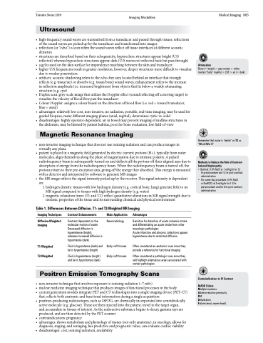
Table 1. Differences Between Diffusion, T1- and T2-Weighted MR Imaging
Medical Imaging MI3
Attenuation
Bone (= bright) > grey matter > white matter (“fatty” myelin) > CSF > air (= dark)
Remember that water is “white” on T2 as “World War II”
Methods to Reduce the Risk of Contrast- Induced Nephropathy
• Optimal: 0.9% NaCl at 1 ml/kg/hr for 12
hr pre-procedure and 12 hr post-contrast
administration
• For same-day procedure: 0.9% NaCl
or NaHCO3 at 3 ml/kg/hr for 1-3 hr pre-procedure and for 6 hr post-contrast administration
Imaging Techniques
Diffusion-Weighted Imaging
T1-Weighted T2-Weighted
Contrast Enhancements
Contrast dependent on the molecular motion of water Decreased diffusion is hyperintense (bright), whereas increased diffusion is hypointense (dark)
Fluid is hypointense (dark) and fat is hyperintense (bright)
Fluid is hyperintense (bright) and fat is hypointense (dark)
Main Application
Neuroradiology
Body soft tissues Body soft tissues
Advantages
Sensitive for detection of acute ischemic stroke and differentiating an acute stroke from other neurologic pathologies
Acute infarction and abscess collections appear hyperintense due to restricted diffusion
Often considered an anatomic scan since they provide a reference for functional imaging
Often considered a pathologic scan since they will highlight edematous areas associated with certain pathologies
Positron Emission Tomography Scans
• non-invasivetechniquethatinvolvesexposuretoionizingradiation(~7mSv)
• nuclearmedicineimagingtechniquethatproducesimagesoffunctionalprocessesinthebody
• current generation models integrate PET and CT technologies into a single imaging device (PET-CT)
that collects both anatomic and functional information during a single acquisition
• positron-producingradioisotopes,suchas18FDG,arechemicallyincorporatedintoametabolically
active molecule (e.g. glucose). These are then injected into the patient, travel to the target organ, and accumulate in tissues of interest. As the radioactive substance begins to decay, gamma rays are produced, and are then detected by the PET scanner
• contraindications: pregnancy
• advantages:showsmetabolismandphysiologyoftissues(notonlyanatomic);inoncology,allowsfor
diagnosis, staging, and restaging; has predictive and prognostic value; can evaluate cardiac viability
• disadvantages:cost,ionizingradiation,availability
Contraindications to IV Contrast
MADD Failure
Multiple myeloma
Adverse reaction previously DM
Dehydration
Failure (renal, severe heart)
