Page 676 - TNFlipTest
P. 676
 MI8 Medical Imaging
Chest Imaging
Toronto Notes 2019
Table 5. Characteristics of Benign and Malignant Pulmonary Nodules
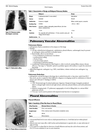
   Figure 10. Pulmonary nodule: bronchogenic carcinoma
Margin Contour Calcification Doubling Time Other Features
Size Cavitation
Satellite Lesions
Malignant
Ill-defined/spiculated (“corona radiata”) Lobulated
Eccentric or stippled
20-460 d
Cavitation, collapse, adenopathy, pleural effusion, lytic bone lesions, smoking history
>3 cm
Yes, especially with wall thickness >15 mm, eccentric cavity, and shaggy internal margins
No
Benign
Well-defined
Smooth
Diffuse, central, popcorn, concentric <20 d or >460 d
<3 cm No
Yes
   Figure 11. Peribronchial cuffing
Pulmonary Vascular Abnormalities
Pulmonary Edema
• pathogenesis:fluidaccumulationintheairspacesofthelungs
• findings
■ vascular redistribution/enlargement, cephalization, pleural effusion, cardiomegaly (may be present in cardiogenic edema and fluid overloaded states)
■ fluid initially collects in interstitium
◆ loss of definition of pulmonary vasculature ◆ peribronchial cuffing
◆ Kerley B lines
◆ reticulonodular pattern
◆ thickening of interlobar fissures
■ as pulmonary edema progresses, fluid begins to collect in alveoli causing diffuse airspace disease often in a “bat wing” or “butterfly” pattern in perihilar regions with tendency to spare the outermost lung fields
• differentialdiagnosis:cardiogenic(e.g.CHF),renalfailure,volumeoverload,non-cardiogenic(e.g. ARDS)
Pulmonary Embolism
• pathogenesis:arterialblockageinthelungsduetoembolifrompelvicorlegveins,rarelyfromPICC lines, ports, air, fat, or amniotic fluid (difficult to diagnose on imaging except by combination of clinical history and CXR and CT findings of ARDS)
• findings
■ CXR: Westermark sign (localized pulmonary oligemia), Hampton’s hump (triangular peripheral
infarct), enlarged right ventricle and right atrium, atelectasis, pleural effusion, and rarely pulmonary
edema
■ definitive imaging study: CT pulmonary angiography to look for filling defect in contrast-filled
pulmonary arteries
■ V/Q scan: Can be used in patients with impaired renal function or in pregnancy
Pleural Abnormalities
Pleural Effusion
Table 6. Sensitivity of Plain Film Views for Pleural Effusion
    X-Ray Projection Lateral decubitus Upright lateral PA
Supine
Minimum Volume to Visualize
25 mL: most sensitive
50 mL: meniscus seen in the posterior costophrenic sulcus 200 mL
Diffuse haziness
    Figure 12. Pleural effusion in lateral view
• ahorizontalfluidlevelisseenonlyinahydropneumothorax(i.e.bothfluidandairwithinpleural cavity)
• effusionmayexertmasseffect,shifttracheaandmediastinumtooppositeside,orcauseatelectasisof adjacent lung
• U/Sissuperiortoplainfilmfordetectionofsmalleffusionsandmayalsoaidinthoracentesis,and POCUS is now standard of care in acute situations


