
Page 682 - TNFlipTest
P. 682
MI14 Medical Imaging
Normal liver appears denser than spleen on CT. If less dense, suspect fatty infiltration
Liver Mass DDx
5Hs
HCC
Hydatid cyst Hemangioma
Hepatic adenoma Hyperplasia (focal nodular)
Abdominal Imaging Toronto Notes 2019 Specific Visceral Organ Imaging
Liver
• U/S:assessmentofcysts,abscesses,tumours,biliarytree
• CT±IV:mostpopularprocedureforimagingtheliverparenchyma(primarylivertumours,metastases,
cysts, abscesses, trauma, cirrhosis)
• MR:alsoexcellentinevaluationofprimarylivertumours,livermetastases,andotherparenchymal
conditions, and is particularly helpful in differentiating common benign hepatic hemangiomas from
primary liver tumours and metastases
• elastography:measuresshearwavevelocitybyU/S(Fibroscan)orMRI(MRelastography)tonon-
invasively quantify liver fibrosis
• findings
■ advanced cirrhosis: liver small and irregular (fibrous scarring, segmental atrophy, regenerating nodules)
■ portal HTN: increased portal vein diameter, collateral veins, splenomegaly (≥12 cm), portal vein thrombosis, recanalization of the umbilical vein
■ porto-systemic shunts: caput medusa, esophageal varices, spontaneous spleno-renal shunt
■ U/S: cirrhosis appears nodular and hyperechoic with irregular areas of atrophy of the right lobe and
hypertrophy of the caudate or left lobes ■ CT: fatty infiltration appears hypodense
• inordertobevisualized,somemassesrequirecontrast
• uponidentifyingaliverlesiononimaging(e.g.U/S),thefollow-upimagingmodalityshouldbeCTor
MR. CT would be four-phase non-contrast, arterial, venous, and delayed to distinguish the common benign liver lesion hemangioma from other tumours
Table 11. Imaging of Liver Masses
Revised Estimates of Diagnostic Test Sensitivity and Specificity in Suspected Biliary Tract Disease
Arch Intern Med 1998;154:2573-2581
Purpose: To assess the sensitivity and specificity of tests used to diagnose cholelithiasis and acute cholecystitis, including ultrasonography (U/S), oral cholecystography, radionucleotide scanning with Technetium, magnetic resonance imaging (MRI) or computed tomography (CT).
Methods: Meta-analysis of studies evaluating the use of different imaging modalities in the diagnosis of biliary tract disease. Main outcomes were sensitivity and specificity of the different imaging modalities, using the gold standard of surgery, autopsy, or 3 mo clinical follow-up for cholelithiasis. For acute cholecystitis, pathologic findings, confirmation of an alternate disease, or clinical resolution during hospitalization for cholecystitis were used as the standard.
Results: Thirty studies were included. For evaluating cholelithiasis, U/S had the best unadjusted sensitivity (0.97; 95% CI 0.95-0.99)
and specificity (0.95, 0.88-1.00) and adjusted (for verification bias) sensitivity (0.84; 0.76-0.92) and specificity (0.99; 95% CI 0.97-1.00). For evaluating acute cholecystitis, radionucleotide scanning has the best sensitivity (0.97; 0.96-0.98) and specificity (0.90; 0.86-0.95).
Conclusion: U/S is the test of choice for diagnosing cholelithiasis and radionucleotide scanning is the superior test for diagnosing acute cholecystitis.
Mass
Benign
Hepatic Adenoma Hemangioma
Focal Nodular Hyperplasia
Abscess Hydatid Cyst
Malignant
HCC Metastases
Spleen
U/S
Most common in young women taking oral contraceptives. Well-defined mass with hyperechoic areas due to hemorrhage
Homogeneous hyperechoic mass
Well-defined mass, central scar seen in 50% Ill-defined, irregular margin, hypoechoic contents Simple/multiloculated cyst
Single/multiple masses, or diffuse infiltration Multiple masses of variable echotexture
CT
Well-defined hypervascular lesion with enlarged central vessel becoming slightly isoattenuating in venous phase
Peripheral globular enhancement in arterial phase scans; central filling and persistent enhancement on delayed scans
Hypervascular mass in arterial phase and isoattenuation to liver in portal venous phase
Low attenuation lesion with an irregular enhancing wall
Low attenuation simple or multiloculated cyst; calcification
Hypervascular; enhances in arterial and washes out in venous phase with portal venous tumour thrombus
Usually low attenuation on contrast-enhanced scan
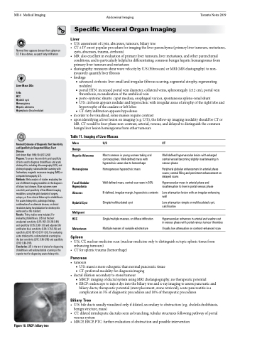
Figure 18. ERCP: biliary tree
• U/S,CT,nuclearmedicinescan(nuclearmedicineonlytodistinguishectopicsplenictissuefrom enhancing tumours)
• CTforsplenictrauma(hemorrhage)
Pancreas
• tumours
■ U/S: mass is more echogenic than normal pancreatic tissue ■ CT: preferred modality for diagnosis/staging
• ductaldilationsecondarytostone/tumour
■ MRCP: imaging of ductal system using MRI cholangiography; no therapeutic potential
■ ERCP: endoscope to inject dye into the biliary tree and x-ray imaging to assess pancreatic and
biliary ducts; therapeutic potential (stent placement, stone retrieval); acute pancreatitis is a complication in 5% of diagnostic procedures and 10% of therapeutic procedures
Biliary Tree
• U/S:bileductsusuallyvisualizedonlyifdilated,secondarytoobstruction(e.g.choledocholithiasis, benign stricture, mass)
• CT:dilatedintrahepaticductulesseenasbranching,tubularstructuresfollowingpathwayofportal venous system
• MRCP,ERCP,PTC:furtherevaluationofobstructionandpossibleintervention
