
Page 684 - TNFlipTest
P. 684
MI16 Medical Imaging
Genitourinary System and Adrenal
Toronto Notes 2019
Angiography requires active blood loss
1-1.5 mL/min under optimal conditions for a bleeding site to be visualized in cases of lower GI bleeding
Imaging Modality Based on Presentation
• Acute testicular pain = Doppler, U/S • Amenorrhea = U/S, MRI (brain)
• Bloating = U/S, CT
• Flank pain = U/S, CT
• Hematuria = U/S, Cystoscopy, CT • Infertility = HSG, MRI
• Lower abdominal mass = U/S, CT • Lower abdominal pain = U/S, CT • Renal colic = U/S, KUB, CT
• Testicular mass = U/S
• Urethral stricture = Urethrogram
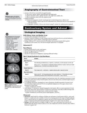
Figure 21. Triphasic CT of an angiomyolipoma: showing fat density with non-contrast scan, mildly enhancing with contrast
Angiography of Gastrointestinal Tract
• anatomyoftheGItractarterialbloodsupplybranches
■ celiac artery: hepatic, splenic, gastroduodenal, left/right gastric
■ superior mesenteric artery: jejunal, ileal, ileo-colic, right colic, middle colic ■ inferior mesenteric artery: left colic, superior rectal
• imagingmodalities
■ conventional angiogram: invasive (usual approach via femoral puncture), catheter used
◆ flush aortography: catheter injection into abdominal aorta, followed by selective arteriography of individual vessels
■ CT angiogram: modality of choice, non-invasive using IV contrast (no catheterization required)
Genitourinary System and Adrenal
Urological Imaging
KUB (Kidney, Ureter, and Bladder X-ray)
• afrontalsupineradiographoftheabdomen
• indication:usefulinevaluationofradio-opaquerenalstones(allstonesbuturicacidandindinavir),
indwelling ureteric stents/catheters, and foreign bodies in abdomen
• findings:additionofIVcontrastexcretedbythekidney(intravenousurogram)allowsgreater
visualization of the urinary tract, but has been largely replaced by CT urography
Abdominal CT
Renal Masses
• Bosniakclassificationforcysticrenalmasses
• classI-II:benignandcanbedisregarded
• classIIF:shouldbefollowed
• classIII-IV:suspiciousformalignancy,requiringadditionalworkup
Table 12. Bozniak Classification for Cystic Renal Masses
NON CONTRAST
ARTERIAL
VENOUS
Classes
Simple Renal Cysts
Class I Class II
Complex Renal Cysts
Class III
Renal Cell Carcinoma
Class IV
Definition
Fluid-attenuating well-defined lesion, no septation, no calcification, no solid components, hair thin wall
Same as class I + fine calcification or moderately thickened calcification in septae or walls; also includes hyperdense cysts (<3 cm) that do not enhance with contrast
Thick irregular walls ± calcifications ± septated, enhancing walls or septa with contrast
Same as class III + soft tissue enhancement with contrast (defined as >10 Hounsfield unit increase, characterizing vascularity) with de-enhancement in venous phase ± areas of necrosis
• plainCTKUBindications:generalimagingofrenalanatomy,renalcolicsymptoms,assessmentofrenal calculi (size and location) and potential sequalae (infection and obstruction), and hydronephrosis prior to urological treatment
• CTurographyindications:investigationofcauseofhematuria,detailedassessmentofurinarytracts (excretory phase), high sensitivity (95%) for uroepithelial malignancies of the upper urinary tracts, assessment of renal calculi
■ phases: unenhanced, excretory
• renaltriphasicCTindications:standardimagingforrenalmasses,allowsaccurateassessmentofrenal
arteries and veins, better characterization of suspicious renal masses, especially in differentiating renal cell carcinoma from more benign masses, and pre-operative staging
■ phases: unenhanced, arterial and venous (nephrographic), excretory
Ultrasound
• indications:initialstudyforevaluationofkidneysizeandnatureofrenalmasses(solidvs.cysticrenal masses vs. complicated cysts); modality of choice for screening patients with suspected hydronephrosis (no IV contrast injection, no radiation to patient, and can be used in patients with renal failure); TRUS useful to evaluate prostate gland and guide biopsies; Doppler U/S to assess renal vasculature
• findings:solidrenalmassesareechogenic(brightonU/S),cysticrenalmasseshavesmoothwell- defined walls with anechoic interior (dark on U/S), and complicated cysts have internal echoes within a thickened, irregular wall
