
Page 683 - TNFlipTest
P. 683
Toronto Notes 2019 Abdominal Imaging “itis” Imaging
Acute Cholecystitis
• pathogenesis:inflammationofgallbladderresultingfromsustainedgallstoneimpactionin cystic duct or, in the case of acalculous cholechystitis, due to gallbladder ischemia or cholestasis (see General Surgery, GS47)
• bestimagingmodality:U/S(bestsensitivityandspecificity);nuclearmedicine(HIDAscan)canhelp diagnose cases of acalculous or chronic cholecystitis
• findings:mostsensitivefindingsarepresenceofgallstonesandpositivesonographicMurphy’ssign (tenderness from pressure of U/S probe over visualized gallbladder). Secondary findings include thickened gallbladder wall (>3 mm), dilated gallbladder and pericholecystic fluid
• management:admit,NPO,IVF,analgesia,cefazolin,earlylaparoscopiccholecystectomy
Acute Appendicitis
• pathogenesis:luminalobstruction→bacterialovergrowth→inflammation/swelling→increased pressure → localized ischemia → gangrene/perforation → localized abscess or peritonitis (see General Surgery, GS27)
• bestimagingmodality:U/SorCT
• findings
■ U/S: thick-walled appendix, appendicolith, dilated fluid-filled appendix, non-compressible; may also demonstrate other causes of RLQ pain (e.g. ovarian abscess, IBD, ectopic pregnancy)
■ CT: enlargement of appendix (>6 mm in outer diameter), enhancement of appendiceal wall, adjacent inflammatory stranding, appendicolith; also facilitates percutaneous abscess drainage
• management:admit,NPO,IVF,analgesia,cefazolin+metronidazole,appendectomy
Acute Diverticulitis
• pathogenesis:erosionoftheintestinalwall(mostcommonlyrectosigmoid)byincreasedintraluminal pressure or inspissated food particles → inflammation and focal necrosis → micro- or macroscopic perforation (see General Surgery, GS31)
• bestimagingmodality:CT,althoughU/Sissometimesused
• contrast:oralandrectalcontrastgivenbeforeCTtoopacifybowel
• findings
■ cardinal signs: thickened wall, mesenteric infiltration, gas-filled diverticula, abscess
■ CT can be used for percutaneous abscess drainage before or in lieu of surgical intervention
■ sometimes difficult to distinguish from perforated cancer (therefore send abscess fluid for cytology
and follow-up with colonoscopy)
■ if chronic, may see fistula (most common to bladder) or sinus tract (linear or branching structures)
• management: ranges from antibiotic treatment to surgical intervention; can use imaging to follow progression
Acute Pancreatitis
• pathogenesis:activationofproteolyticenzymeswithinpancreaticcellsleadingtolocal
and systemic inflammatory response (see Gastroenterology, G44); a clinical/biochemical diagnosis
• best imaging modality: imaging used to support diagnosis and evaluate for complications (diagnosis cannot be excluded by imaging alone)
■ U/S good for screening and follow-up
■ CT is useful in advanced stages and in assessing for complications (1st line imaging test)
• findings
■ U/S: hypoechoic enlarged pancreas (if ileus present, gas obscures pancreas)
■ CT: enlarged pancreas, edema, stranding changes in surrounding fat with indistinct fat planes,
mesenteric and Gerota’s fascia thickening, pseudocyst in lesser sac, abscess (gas or thick-walled fluid collection), pancreatic necrosis (low attenuation gas-containing non-enhancing pancreatic tissue), hemorrhage
• management:supportivetherapy
■ CT-guided needle aspiration and/or drainage done for abscess when clinically indicated ■ pseudocyst may be followed by CT and drained if symptomatic
Chronic Pancreatitis
• pathogenesis:(seeGastroenterology,G45)
• bestimagingmodality:MRCP(canshowcalcificationandductobstruction)
• findings:U/S,CTscan,andMRImayshowcalcifications,ductaldilatation,enlargementofthepancreas
and fluid collections (e.g. pseudocysts) adjacent to the gland
Medical Imaging MI15
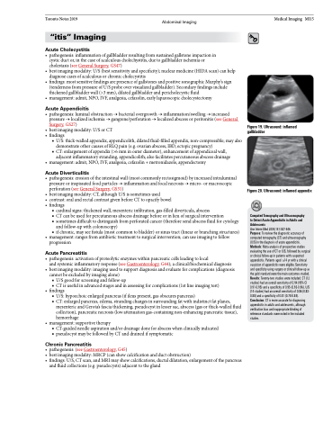
Figure 19. Ultrasound: inflamed gallbladder
Figure 20. Ultrasound: inflamed appendix
Computed Tomography and Ultrasonography to Detect Acute Appendicitis in Adults and Adolescents
Ann Intern Med 2004;141:537-546
Purpose: To review the diagnostic accuracy of computed tomography (CT) and ultrasonography (U/S) in the diagnosis of acute appendicitis. Methods: Meta-analysis of prospective studies evaluating the use of CT or U/S, followed by surgical or clinical follow-up in patients with suspected appendicitis.Patientsaged ≥14yrwithaclinical suspicion of appendicitis were eligible. Sensitivity and specificity using surgery or clinical follow-up as the gold standard were the main outcomes studied. Results: Twenty-two studies were included. CT (12 studies) had an overall sensitivity of 0.94 (95% CI 0.91-0.95) and a specificity of 0.95 (0.93-0.96). U/S (14 studies) had an overall sensitivity of 0.86 (0.83- 0.88) and a specificity of 0.81 (0.78-0.84). Conclusion: CT is more accurate for diagnosing appendicitis in adults and adolescents, although verification bias and inappropriate blinding of reference standards were noted in the included studies.
