
Page 686 - TNFlipTest
P. 686
MI18 Medical Imaging
Neuroradiology
Toronto Notes 2019
Table 14. Typical and Atypical Findings on a Sonohysterogram
Finding
Polyps Leiomyoma
Hyperplasia and Cancer
Adhesions
Typical
A well-defined, homogeneous, polypoid lesion isoechoic to the endometrium with preservation of the endometrial- myometrial interface
Well-defined, broad-based, hypoechoic, solid masses with shadowing. Overlying layer of endometrium is echogenic and distorts the endometrial-myometrial interface
Diffuse echogenic endometrial thickening without focal abnormality, although focal lesions can occur. Endometrial cancer is typically a diffuse process, but early cases can be focal and appear as a polypoid mass
Mobile, thin, echogenic bands that cut across the endometrial cavity
Atypical
Cystic components, multiple polyps, broad base, hypoechogenicity or heterogeneity
Pedunculation or multilobulated surface
Thick, broad-based bands that can completely obliterate the endometrial cavity, as in Asherman’s syndrome
Modality Based on Neuropathology Presentation
• Cognitive decline = CT
• Cord compression = MRI
• Decreased level of consciousness = CT
• Fish bone/other swallowed foreign body =
CT
• Low back pain, radiculopathy = MRI
• Multiple sclerosis = MRI
• Neck infection = CT
• Orbital infection = CT
• Rule out bleed = CT
• Rule out aneurysm = CTA, MRA
• Seizure = CT
• Sinusitis = CT
• Stroke = CT, MRI
• Trauma = CT
• Weakness, systemically unwell = CT
Figure 26. Epidural hematoma
Figure 27. Subdural hematoma
Figure 28. Subarachnoid hemorrhage
CT
• indications:excellentstudyforevaluationofbonyandintracranialabnormalities
• oftendonefirstwithoutandthenwithIVcontrasttoshowvascularstructuresoranomalies
Adrenal Mass
• imagingmodality:mostoftenidentifiedonCTscanas‘incidentaloma,’canalsouseCT/MRIto distinguish benign from malignant masses
Table 15. Adrenal Mass Findings on CT and MRI
Factors
Diameter (CT) Shape (CT)
Texture (CT)
Vascularity (CT)
Washout of Contrast Medium on CT
Growth
Other Findings
MRI on T2 Weighted Imaging
Adrenocortical Adenoma
Usually ≤3 cm Smooth margins and
round/oval Homogeneous
Not highly vascular ≥50% at 10 min
Stable or very slow (<1 cm/yr)
Usually low density due to intracellular fat
Isointense in relation to liver
Adrenocortical Carcinoma
Usually ≥4 cm Irregular with unclear
margins
Heterogeneous with mixed densities
Usually vascular <50% at 10 min
Usually rapid (>2 cm/yr)
Necrosis, calcifications, and hemorrhage
Hyperintense in relation to liver
Pheochromocytoma
Usually >3 cm
Round/oval with clear margins
Heterogeneous with cystic areas
Usually vascular <50% at 10 min
Slow (0.5-1 cm/yr) Hemorrhage
Markedly hyperintense in relation to liver
Metastasis
Variable around <3 cm
Oval/irregular with unclear margins
Heterogeneous with mixed densities
Usually vascular <50% at 10 min
Variable
Occasionally hemorrhage
Hyperintense in relation to liver
Neuroradiology
Modalities
• CTisthemodalityofchoiceformostneuropathology;evenundercircumstanceswhereMRIis preferred
• CTisfrequentlytheinitialstudyperformedbecauseofitsspeed,availability,andlowercost
■ acute craniofacial trauma: CT is best for visualizing “bone and blood”; MRI is used only when CT
fails to detect an abnormality despite strong clinical suspicion
■ acute stroke: MRI ideal, CT most frequently used
■ acute headache with focal neurologic signs
■ suspected subarachnoid or intracranial hemorrhage
■ suspected hydrocephalus
■ meningitis: rule out mass effect (e.g. cerebral herniation, shift) prior to lumbar puncture
■ tinnitus and vertigo: CT and MRI are used in combination to detect bony abnormalities and CN
VIII tumours, respectively
Skull Films
• rarelyperformed,generallynotindicatedfornon-penetratingheadtrauma
• indications:screeningfordestructivebonylesions(e.g.metastases),metabolicdisease,skullanomalies,
post-operative changes and confirmation of hardware placement, skeletal surveys, multiple myeloma
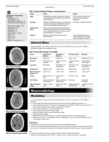
Figure 29. Intraparenchymal hemorrhage
