
Page 687 - TNFlipTest
P. 687
Toronto Notes 2019 Neuroradiology Medical Imaging MI19
• vascularstructuresandareasofblood-brainbarrierimpairmentareopaque(e.g.hyperattenuatingor bright/show enhancement) with contrast injection
■ when in doubt, look for Circle of Willis or confluence of sinuses to determine presence of contrast enhancement
• posteriorfossacanbeobscuredbyextensivebony-relatedstreakartifact
• ruleoutskullfracture,epiduralhematoma(lenticularshape),subduralhematoma(crescenticshape),
subarachnoid hemorrhage, space occupying lesion, hydrocephalus, and cerebral edema
• multiplanarimagingcanbeperformedwithnewergenerationofmultidetectorCTscanners
Myelography
• introductionofwater-soluble,low-osmoticcontrastmediaintosubarachnoidspaceusinglumbar puncture followed by x-ray
• LargelyreplacedbyMRIorCTmyelogram
• indications:excellentstudyfordischerniation,traumaticnerverootavulsion,patientswith
contraindication to MRI
MRI
• indications:finerneuroanatomicdefinition,bettergrey-whitematterdifferentiation(especiallyT1- weighted series), better evaluation of edema extent (better tumour detection), allows evaluation of structures obscured by bony artifacts on CT (posterior fossa structures), multiplanar imaging helpful in pre-operative assessment
Cerebral Angiography/CT Angiography/MR Angiography
• indications:evaluationofvascularlesionssuchasatheroscleroticdisease,aneurysms,vascular malformations, arterial dissection
• conventionaldigitalsubtractionangiographyremainsthegoldstandardfortheassessmentofneckand intracranial vessels; however, it is an invasive procedure requiring arterial (femoral) puncture; catheter manipulation confers risk of vessel injury (e.g. dissection, occlusion, vasospasm, emboli)
• MRAmethods(phasecontrast,timeofflight,gadolinium-enhanced)andCTAaremuchlessinvasive without risk to intracranial or neck vessels
• MRAandCTAareoftenusedfirstas‘screeningtests’fortheassessmentofsubarachnoidhemorrhage, vasospasm, or aneurysms
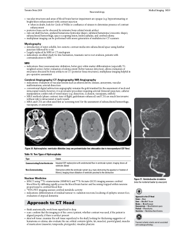
Figure 30. Hydrocephalus: ventricular dilatation (may see periventricular low attenuation due to transependymal CSF flow) Table 16. Two Types of Hydrocephalus
Type
Communicating/Extra-Ventricular Non-Communicating
Nuclear Medicine
Cause
Impaired CSF reabsorption with unobstructed flow in ventricular system; imaging shows all ventricles dilated
Obstruction within the ventricular system (e.g. mass obstructing the aqueduct or foramen of Monro); imaging shows dilatation of ventricles proximal to the obstruction
• SPECTusing99mTc-exametazime(HMPAO)and99mTc-bicisate(ECD)imagingassessescerebral blood flow by diffusing rapidly across the blood brain barrier and becoming trapped within neurons proportional to cerebral blood flow
• 18FDGPETimagingassessescerebralmetabolicactivity
• indications:differentiationofresidualtumourvs.radiationnecrosis;localizingofepilepticseizurefoci;
evaluation of atypical dementia
Approach to CT Head
• thinkanatomically,workfromsuperficialtodeep
• scan:confirmthattheimagingisofthecorrectpatient,whethercontrastwasused,ifthepatientis
aligned properly, if there is artifact present
• skin/softtissue:examinethesofttissuesuperficialtotheskull,lookingforthickeningsuggestiveof
hematoma or edema; also evaluate the ear, orbital contents (globe, fat, muscles), parotid gland, muscles of mastication (masseter, temporalis, pterygoids), visualize pharynx
Figure 31. Vertebrobasilar circulation
(note the incidental basilar tip aneurysm)
Approach to the CT Head
Some = Scan
Sore = Skin/Soft Tissue
Brains = Bone/Airspace Demonstrate = Dura/Subdural space Pushed = Parenchyma
Ventricles = Ventricles/Sulci/Cisterns
Transient ischemic attacks are not associated with radiological findings
