
Page 689 - TNFlipTest
P. 689
Toronto Notes 2019 Musculoskeletal System Medical Imaging MI21
■ perivascular and interstitial edema may be prominent ■ spinal cord lesions typical of MS
◆ little or no cord swelling
◆ unequivocal hyperintensity on T2-weighted sequences
◆ size at least 3 mm but less than 2 vertebral segments in length
◆ occupy only part of the cord in cross-section
◆ focal (i.e. clearly delineated and circumscribed on T2-weighted sequences)
CNS Infections
• meningitis
■ pathogenesis: inflammation of the pia or arachnoid mater, most often secondary to hematogenous
spread from infection or via organisms gaining access across areas not protected by the blood-brain
barrier (choroid plexus or circumventricular organs)
• pathogensinclude:S.pneumoniae,H.influenzae,N.meningitidis,L.monocytogenes
■ best imaging modality: MRI (T2-weighted/FLAIR) ■ findings
◆ meningeal enhancement (following the gyri/sulci and/or basal cisterns), hydrocephalus (communicating), cerebral swelling, subdural effusion
• anormalMRIdoesnotruleoutleptomeningitis
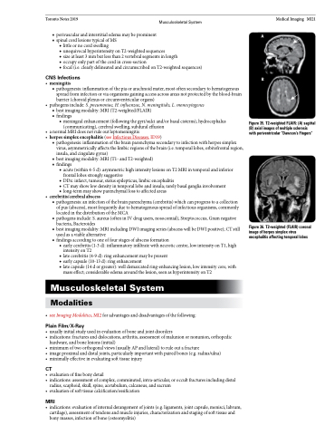
• herpessimplexencephalitis(seeInfectiousDiseases,ID19)
■ pathogenesis: inflammation of the brain parenchyma secondary to infection with herpes simplex virus, asymmetrically affects the limbic regions of the brain (i.e. temporal lobes, orbitofrontal region, insula, and cingulate gyrus)
■ best imaging modality: MRI (T1- and T2-weighted)
■ findings
◆ acute (within 4-5 d): asymmetric high intensity lesions on T2 MRI in temporal and inferior frontal lobes strongly suggestive
◆ DDx: infarct, tumour, status epilepticus, limbic encephalitis
◆ CT may show low density in temporal lobe and insula; rarely basal ganglia involvement ◆ long-term may show parenchymal loss to affected areas
• cerebritis/cerebralabscess
■ pathogenesis: an infection of the brain parenchyma (cerebritis) which can progress to a collection
of pus (abscess), most frequently due to hematogenous spread of infectious organisms, commonly
located in the distribution of the MCA
■ pathogens include: S. aureus (often in IV drug users, nosocomial), Streptococcus, Gram negative
bacteria, Bacteroides
■ best imaging modality: MRI including DWI imaging series (abscess will be DWI positive); CT still
used as a viable alternative
■ findings according to one of four stages of abscess formation
◆ early cerebritis (1-3 d): inflammatory infiltrate with necrotic centre, low intensity on T1, high intensity on T2
◆ late cerebritis (4-9 d): ring enhancement may be present
◆ early capsule (10-13 d): ring enhancement
◆ late capsule (14 d or greater): well demarcated ring-enhancing lesion, low intensity core, with
mass effect; considerable edema around the lesion, seen as hyperintensity on T2
Musculoskeletal System
Modalities
• seeImagingModalities,MI2foradvantagesanddisadvantagesofthefollowing:
Plain Film/X-Ray
• usuallyinitialstudyusedinevaluationofboneandjointdisorders
• indications:fracturesanddislocations,arthritis,assessmentofmalunionornonunion,orthopedic
hardware, and bone lesions (initial)
• minimumoftwoorthogonalviews(usuallyAPandlateral)toruleoutafracture
• imageproximalanddistaljoints,particularlyimportantwithpairedbones(e.g.radius/ulna)
• minimallyeffectiveinevaluatingsofttissueinjury
CT
• evaluationoffinebonydetail
• indications:assessmentofcomplex,comminuted,intra-articular,oroccultfracturesincludingdistal
radius, scaphoid, skull, spine, acetabulum, calcaneus, and sacrum
• evaluationofsofttissuecalcification/ossification
MRI
• indications:evaluationofinternalderangementofjoints(e.g.ligaments,jointcapsule,menisci,labrum, cartilage), assessment of tendons and muscle injuries, characterization and staging of soft tissue and bony masses, infection of bone (osteomyelitis)
Figure 35. T2-weighted FLAIR: (A) sagittal (B) axial images of multiple sclerosis with periventricular “Dawson’s Fingers”
Figure 36. T2-weighted (FLAIR) coronal image of herpes simplex virus encephalitis affecting temporal lobes
A
B
