
Page 73 - TNFlipTest
P. 73
Toronto Notes 2019 Cardiac Diagnostic Tests Generalized Edema
• increasedhydrostaticpressure/fluidoverload
■ heart failure, pregnancy, drugs (e.g. CCBs), iatrogenic (e.g. IV fluids)
• decreasedoncoticpressure/hypoalbuminemia
■ liver cirrhosis, nephrotic syndrome, malnutrition
• increasedinterstitialoncoticpressure ■ myxedema
• increasedcapillarypermeability ■ severe sepsis
• hormonal
■ hypothyroidism, exogenous steroids, pregnancy, estrogens
Palpitations
• anunpleasantsensationofcardiacactivity
• palpitationsthatarecontinuousrapidheartaction:
■ conditions causing sinus tachycardia - endocrine (thyrotoxicosis, pheochromocytoma, and hypoglycemia), systemic (anemia, fever), drugs (stimulants and anticholinergics), and psychiatric (panic attacks)
■ conditions causing pathologic tachycardia: SVT (atrial tach, A fib, and A flutter); re-entrant SVT, VT
• palpitationswithirregular/intermittentsensations • PACs, PVCs
Dyspnea
• cardiovascular
■ due to elevated pulmonary venous pressure – acute MI, CHF/LV failure, aortic/mitral stenosis,
aortic/mitral regurgitation, arrhythmia, cardiac tamponade, constrictive pericarditis, and left-sided
obstructive lesions (e.g. left atrial myxoma) • respiratory
■ airway disease
◆ asthma, COPD exacerbation, and upper airway obstruction (anaphylaxis, foreign body, and
mucus plugging)
■ parenchymal lung disease
◆ ARDS, pneumonia, interstitial lung disease ■ pulmonary vascular disease
◆ PE, pulmonary HTN, pulmonary vasculitis ■ pleural disease
◆ pneumothorax, pleural effusion • neuromuscularandchestwalldisorders
■ C-spine injury
◆ polymyositis, myasthenia gravis, Guillain-Barré syndrome, and kyphoscoliosis
• anxiety/psychosomatic
• hematological/metabolic
■ anemia, acidosis, hypercapnia • drugsandpoisons
■ CNS depressants, carbon monoxide poisoning
Cardiac Diagnostic Tests
Electrocardiography Basics
Description
• agraphicalrepresentation(timeversusamplitudeofelectricalvectorprojection)oftheelectricalactivity of the heart
• ontheECGgraph
■ the horizontal axis represents time (at usual paper speed 25 mm/s)
◆ 1 mm (1 small square) = 40 msec
◆ 5 mm (1 large square) = 200 msec
■ theverticalaxisrepresentsvoltage(atusualstandardgainsetting10mm/mV)
◆ 1 mm (1 small square) = 0.1 mV ◆ 10 mm (2 large squares) = 1 mV
Cardiology and Cardiac Surgery
C5
RA
LA
V2V3
RL
LL
V1
V4 V5 V6
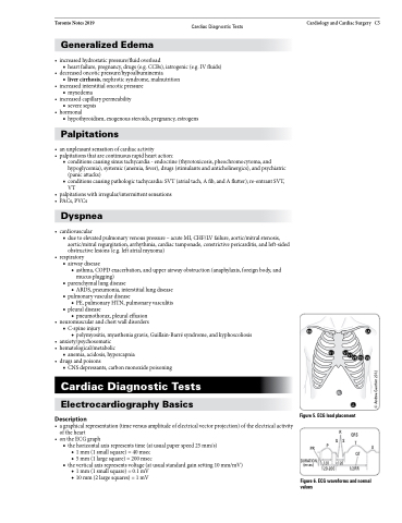
Figure 5. ECG lead placement
PR
DURATION (msec)
P
120 <120 120-200
U
R QRS Q S T
QT 1/2RR
Figure 6. ECG waveforms and normal values
© Andrea Gauthier 2012
