
Page 75 - TNFlipTest
P. 75
Toronto Notes 2019
Approach to ECGs
Cardiology and Cardiac Surgery C7
Table 1. Conduction Abnormalities
Left Bundle Branch Block (LBBB)
Complete LBBB
QRS duration >120 msec
Broad notched R waves in leads V4, and V5, and usually I, aVL Deep broad S waves in leads V1-2
Secondary ST-T changes (-ve in leads with broad notched R waves, +ve in V1-2) are usually present LBBBcanmaskECGsignsofMI
LBBB: lead V 1 negative, V6 positive and notched
Right Bundle Branch Block (RBBB)
Complete RBBB
QRS duration >120 msec
Positive QRS in lead V1 (rSR’ or occasionally broad R wave) Broad S waves in leads I, V5-6 (>40 msec)
Usually secondary T wave inversion in leads V1-2
Frontal axis determination using only the first 60 msec RBBB:V1ispositive(rSR’), V6haslateSwave
Left Bundle Branch Block
Right Bundle Branch Block
Left Ventricular Hypertrophy
Right Ventricular Hypertrophy
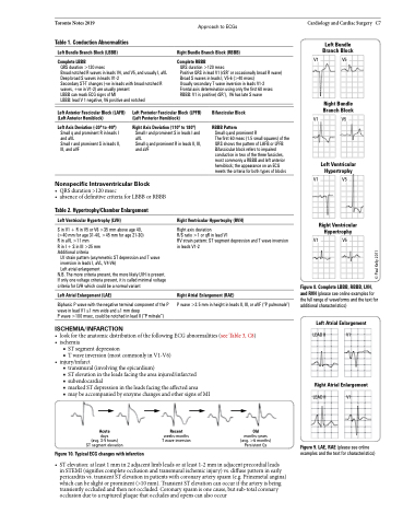
Figure 8. Complete LBBB, RBBB, LVH, and RVH (please see online examples for the full range of waveforms and the text for additional characteristics)
Left Atrial Enlargement
LEAD II
Right Atrial Enlargement
LEAD II
Figure 9. LAE, RAE (please see online examples and the text for characteristics)
V1
V5
Left Anterior Fascicular Block (LAFB) (Left Anterior Hemiblock)
Left Axis Deviation (-30o to -90o)
Small q and prominent R in leads I and aVL
Small r and prominent S in leads II, III, and aVF
Left Posterior Fascicular Block (LPFB) (Left Posterior Hemiblock)
Right Axis Deviation (110o to 180o)
Small r and prominent S in leads I and aVL
Small q and prominent R in leads II, III, and aVF
Bifascicular Block
RBBB Pattern
Small q and prominent R
The first 60 msec (1.5 small squares) of the QRS shows the pattern of LAFB or LPFB Bifascicular block refers to impaired conduction in two of the three fascicles, most commonly a RBBB and left anterior hemiblock; the appearance on an ECG meets the criteria for both types of blocks
V1
V5
V1
V5
Nonspecific Intraventricular Block
• QRSduration>120msec
• absenceofdefinitivecriteriaforLBBBorRBBB
Table 2. Hypertrophy/Chamber Enlargement
Left Ventricular Hypertrophy (LVH)
S in V1 + R in V5 or V6 >35 mm above age 40, (>40 mm for age 31-40, >45 mm for age 21-30) R in aVL >11 mm
R in I + S in III >25 mm
Additional criteria
LV strain pattern (asymmetric ST depression and T wave inversion in leads I, aVL, V4-V6)
Left atrial enlargement
N.B. The more criteria present, the more likely LVH is present. If only one voltage criteria present, it is called minimal voltage criteria for LVH which could be a normal variant
Left Atrial Enlargement (LAE)
Biphasic P wave with the negative terminal component of the P wave in lead V1 ≥1 mm wide and ≥1 mm deep
P wave >100 msec, could be notched in lead II (“P mitrale”)
ISCHEMIA/INFARCTION
Right Ventricular Hypertrophy (RVH)
Right axis deviation
R/S ratio >1 or qR in lead V1
RV strain pattern: ST segment depression and T wave inversion in leads V1-2
Right Atrial Enlargement (RAE)
P wave >2.5 mm in height in leads II, III, or aVF (“P pulmonale”)
V1
V5
V1
• lookfortheanatomicdistributionofthefollowingECGabnormalities(seeTable3,C8) • ischemia
■ ST segment depression
■ T wave inversion (most commonly in V1-V6) • injury/infarct
■ transmural (involving the epicardium)
■ ST elevation in the leads facing the area injured/infarcted
■ subendocardial
■ marked ST depression in the leads facing the affected area
■ may be accompanied by enzyme changes and other signs of MI
V1
Acute
days
(avg. 3-5 hours) ST segment elevation
Figure 10. Typical ECG changes with infarction
Recent
weeks-months T wave inversion
Old
months-years (avg. >6 months) Persistent Qs
• STelevation:atleast1mmin2adjacentlimbleadsoratleast1-2mminadjacentprecordialleads
in STEMI (signifies complete occlusion and transmural ischemic injury) vs. diffuse pattern in early pericarditis vs. transient ST elevation in patients with coronary artery spasm (e.g. Prinzmetal angina) which can be slight or prominent (>10 mm). Transient ST elevation can occur if the artery is being transiently occluded and then not occluded. Coronary spasm is one cause, but sub-total coronary occlusion due to a ruptured plaque that occludes and opens can also occur
© Paul Kelly 2011
