
Page 71 - TNFlipTest
P. 71
Toronto Notes 2019
Basic Anatomy Review
Cardiology and Cardiac Surgery C3
120
60
0 120
80 40
ECG
Heart Sounds
AV opens
MV closes
Aortic pressure
Carotid Pulse
JVP Waveform
Systole
Diastole AV closes
LV pressure MV opens
Legend:
AV – aortic valve LA – left atrium LV – left ventricle MV – mitral valve
LA Pressure
a
x c
x’
S1 S2
v
a wave
LV end-diastolic volume
Cardiac Sounds
x descent
y
R QS
S1
T
LV end-systolic volume
0.8
0
0.4 Time (sec)
v wave
y descent
P S4
S2
S3
PT QRS
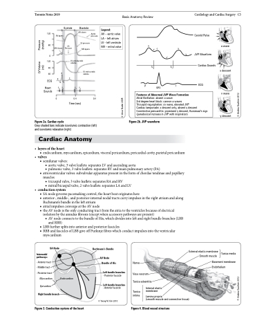
Figure 2b. JVP waveform
ECG
Features of Abnormal JVP Wave Formation
Atrial fibrillation: absent a wave
3rd degree heart block: cannon a waves
Tricuspid regurgitation: cv wave, elevated JVP
Cardiac tamponade: x descent only, absent y descent Constrictive pericarditis: prominent y descent, Kussmaul's sign (paradoxical increase in JVP with inspiration)
Figure 2a. Cardiac cycle
Grey shaded bars indicate isovolumic contraction (left) and isovolumic relaxation (right)
Cardiac Anatomy
• layersoftheheart
■ endocardium,myocardium,epicardium,visceralpericardium,pericardialcavity,parietalpericardium • valves
■ semilunar valves:
◆ aortic valve, 3 valve leaflets: separates LV and ascending aorta
◆ pulmonic valve, 3 valve leaflets: separates RV and main pulmonary artery (PA)
■ atrioventricular valves: subvalvular apparatus present in the form of chordae tendinae and papillary muscles
◆ tricuspid valve, 3 valve leaflets: separates RA and RV
◆ mitral/bicuspid valve, 2 valve leaflets: separates LA and LV
• conductionsystem
■ SA node governs pacemaking control; the heart beat originates here
■ anterior-, middle-, and posterior-internal nodal tracts carry impulses in the right atrium and along
Bachmann’s bundle in the left atrium
■ atrial impulses converge at the AV node
■ the AV node is the only conducting tract from the atria to the ventricles because of electrical
isolation by the annulus fibrosis (except when accessory pathways are present)
◆ AV node connects to the bundle of His, which divides into left and right bundle branches (LBB
and RBB)
■ LBB further splits into anterior and posterior fascicles
■ RBB and fascicles of LBB give off Purkinje fibres which conduct impulses into the ventricular
myocardium
SA Node
Internodal pathways:
Bachmann’s Bundle
AV Node Bundle of His
Left bundle branches
- Posterior fascicle
Left bundle branches
- Anterior fascicle © Young M. Kim 2010
External elastic membrane Smooth muscle
Tunica media
- Anterior tract - Middle tract
- Posterior tract
Myocardium
Epicardium
Right bundle branch
Nerve
Vasa vasorum Tunica adventitia
Basement membrane Endothelium
Endocardium
Tunica intima
Internal elastic membrane
Lamina propria
(smooth muscle and connective tissue)
Figure 3. Conduction system of the heart
Figure 4. Blood vessel structure
© Andrea Gauthier 2012 © Andrea Gauthier 2012
© Anas Nader 2009
LV Volume Pressure (ml) (mmHg)
