
Page 83 - TNFlipTest
P. 83
Toronto Notes 2019 Approach to ECGs
Left Heart Catheterization
• description
■ accomplished by introducing a catheter into the brachial or femoral artery and advancing it through
the aorta, across the aortic valve, and into the left ventricle
■ evaluates mitral and aortic valvular defects and myocardial disease
■ systolic and end-diastolic pressure tracings recorded
■ LV size, wall motion and ejection fraction can be assessed by injecting contrast into the LV (left
ventriculography) via femoral/radial artery catheterization
■ cardiac output (measured by the Fick oxygen method or the indicator dilution method)
• indications
■ identification of the extent and severity of CAD and evaluation of left ventricular function
■ assessment of the severity of valvular or myocardial disorders (e.g. aortic stenosis or insufficiency,
mitral stenosis or insufficiency, and various cardiomyopathies) to determine the need for surgical
correction
■ collection of data to confirm and complement noninvasive studies
■ determination of the presence of CAD in patients with confusing clinical presentations or chest pain
of uncertain origin
• contraindications
■ severe uncontrolled hypertension ■ ventricular arrhythmias
■ acute stroke
■ severe anemia
■ active gastrointestinal bleeding
■ allergy to radiographic contrast
■ acute renal failure
■ uncompensated congestive failure (so that the patient cannot lie flat) ■ unexplained febrile illness or untreated active infection
■ electrolyte abnormalities (e.g. hypokalemia)
■ severe coagulopathy
• risks
■ complications for diagnostic catheterization <1%
■ inadequate diagnostic procedures occur in <1% of cases
■ within 24 h of catheterization: death, MI, or stroke (0.2% to 0.3% of patients)
Coronary Angiography
• description
■ radiographic visualization of the coronary vessels after injection of radiopaque contrast media ■ coronary vasculature accessed via the coronary ostia
• indications
■ to define the coronary anatomy and the degree of luminal obstruction of the coronary arteries
■ to determine the presence and extent of obstructive CAD
■ to assess the feasibility and appropriateness of various forms of therapy, such as revascularization by
percutaneous or surgical interventions
■ can also be used when the diagnosis of CAD is uncertain and CAD cannot be reasonably excluded
by noninvasive techniques
• contraindications:severerenalfailure(duetocontrastagenttoxicity–mustcheckpatient’srenalstatus)
• risks:majorcomplications<2%,butincreasedinpatientswithpre-existingrenalfailure(especiallyin
Cardiology and Cardiac Surgery C15
Chambers Pressure (systolic; mmHg)
Right atrium/central 1-8 venous
Right ventricle 1-8 (15-30) Pulmonary artery 4-12 (15-30)
Left atrium/ pulmonary 4-12 capillary wedge
Left ventricle end diastolic 4-12
diabetic patients)
ACC/AHA 2011 Recommended Indications for Coronary Angiography
• Disabling (CCS classes III and IV) chronic
stable angina despite medical therapy
• High-risk criteria on clinical assessment or
non-invasive testing
• Serious ventricular arrhythmia or CHF • Uncertain diagnosis or prognosis after
non-invasive testing
• Inability to undergo non-invasive testing
Coronary Angiography
Gold standard for localizing and quantifying CAD
Hemodynamically significant stenosis is defined as 70% or more narrowing of the luminal diameter
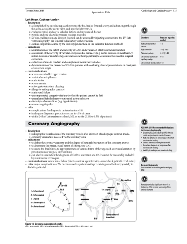
1 - Inferobasal RAO
2 - Inferoapical 5
3 - Apical 1 4 4 - Anteroapical
5 - Anterobasal 2
Figure 18. Coronary angiogram schematic
AM
1st septal LAD
OM1 OM2 OM3
AM = acute marginal; LAD = left anterior descending; OM = obtuse marginal; RCA = right coronary artery
Carotid
Circumflex Int.
AM 3
RCA
1st diag.
2nd diag.
Posterolateral branches
PIV
AV
