
Page 912 - TNFlipTest
P. 912
OP22 Ophthalmology
Retina Toronto Notes 2019
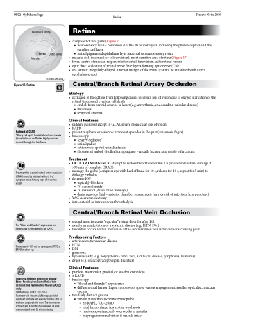
Peripheral retina
Fovea Optic nerve Macula
© Tobi Lam 2012
•
• • • •
Retina
composedoftwoparts(Figure2)
■ neurosensory retina: comprises 9 of the 10 retinal layers, including the photoreceptors and the
ganglion cell layer
■ retinal pigmented epithelium layer: external to neurosensory retina
macula:richincones(forcolourvision);mostsensitiveareaofretina(Figure17) fovea:centreofmacula;responsiblefordetail,finevision,lacksretinalvessels
optic disc: collection of retinal nerve fibre layers forming optic nerve (CN2) oraserrata:irregularly-shaped,anteriormarginoftheretina(cannotbevisualizedwithdirect ophthalmoscope)
Central/Branch Retinal Artery Occlusion
Figure 17. Retina
Hallmark of CRAO
“Cherry-red spot” located at centre of macula (visualization of unaffected highly vascular choroid through the thin fovea)
Treatment for a central retinal artery occlusion (CRAO) must be initiated within 2 h of symptom onset for any hope of restoring vision
The “blood and thunder” appearance on fundoscopyisveryspecificfor CRVO
There is an 8-10% risk of developing CRVO or BRVO in other eye
Intravitreal Aflibercet injection for Macular Edema Resulting from Central Retinal Vein Occlusion: One Year results of Phase 3 GALILEO study
Ophthalmology 2014; 121(1) 202-8
Treatment with intravitreal aflibercept provided significant functional and anatomic benefits after 52 weeks as compared with sham. The improvement achieved after 6 monthly doses at week 24 were maintained until week 52 with prn dosing.
Etiology
• occlusionofbloodflowfromfollowingcausesresultsinlossofvisionduetooxygenstarvationofthe retinal tissues and eventual cell death
■ emboli from carotid arteries or heart (e.g. arrhythmia, endocarditis, valvular disease) ■ thrombus
■ temporal arteritis
Clinical Features
• sudden,painless(exceptinGCA),severemonocularlossofvision
• RAPD
• patientmayhaveexperiencedtransientepisodesinthepast(amaurosisfugax) • fundoscopy
■ “cherry-red spot”
■ retinal pallor
■ cotton wool spots (retinal infarcts)
■ cholesterol emboli (Hollenhorst plaques) – usually located at arteriole bifurcations
Treatment
• OCULAREMERGENCY:attempttorestorebloodflowwithin2h(irreversibleretinaldamageif >90 min of complete CRAO)
• massagetheglobe(compresseyewithheelofhandfor10s,releasefor10s,repeatfor5min)to dislodge embolus
• decreaseIOP
■ topical β-blockers
■ IVacetazolamide
■ IV mannitol (draws fluid from eye)
■ drain aqueous fluid – anterior chamber paracentesis (carries risk of infection, lens puncture)
• YAGlaserembolectomy
• intra-arterialorintra-venousthrombolysis
Central/Branch Retinal Vein Occlusion
• secondmostfrequent“vascular”retinaldisorderafterDR
• usuallyamanifestationofasystemicdisease(e.g.HTN,DM)
• thrombusoccurswithinthelumenofthecentralretinalvein/arteriovenouscrossingpoint
Predisposing Factors
• arterioscleroticvasculardisease • HTN
• DM
• glaucoma
• hyperviscosity(e.g.polycythemiarubravera,sickle-celldisease,lymphoma,leukemia) • drugs(e.g.oralcontraceptivepill,diuretics)
Clinical Features
• painless,monocular,gradual,orsuddenvisionloss • ±RAPD
• fundoscopy
■ “blood and thunder” appearance
■ diffuse retinal hemorrhages, cotton wool spots, venous engorgement, swollen optic disc, macular
edema
• twofairlydistinctgroups
■ venous stasis/non-ischemic retinopathy
◆ no RAPD, VA ~20/80
◆ mild hemorrhage, few cotton wool spots
◆ resolves spontaneously over weeks to months ◆ may regain normal vision if macula intact
