
Page 914 - TNFlipTest
P. 914
OP24 Ophthalmology
Retinitis Pigmentosa Inherited Forms
• Autosomal recessive: most common • Autosomal dominant: best prognosis • X-linked: worst prognosis
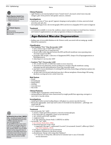
Triad of Retinitis Pigmentosa
APO
Arteriolar narrowing
Perivascular bony-spicule pigmentation Optic disc pallor
Retina Toronto Notes 2019 • nightblindness,decreasedperipheralvision(“tunnelvision”),decreasedcentralvision(macular
changes), glare (from posterior subcapsular cataracts; common)
Investigations
• fundoscopy:areasof“bone-spicule”pigmentclumpinginmid-peripheryofretina,narrowedretinal arterioles, pale optic disc
• electrophysiologicaltests:electroretinography(ERG)andelectrooculography(EOG)assistindiagnosis
Treatment
• notreatmentsavailabletoreversethecondition;cataractextractionimprovesvisualfunction;vitaminA and vitamin E supplementation can reduce progression of disease in some patients
Age-Related Macular Degeneration
• leadingcauseofirreversibleblindnessintheWesternworld,associatedwithincreasingage,usually bilateral but asymmetric
Classification
• Non-Exudative/“Dry”(Non-Neovascular)AMD
■ most common type of AMD (90% of cases)
■ slowly progressive loss of visual function
■ drusen: yellow-white deposits between the RPE and Bruch’s membrane (area separating inner
choroidal vessels from RPE)
■ geographic RPE atrophy: coalescence of depigmented RPE, clumps of focal hyperpigmentation or
hypopigmentation
■ may progress to neovascular AMD
• Exudative/“Wet”(Neovascular)AMD
■ 10% of AMD, but 80% of AMD that results in severe vision loss
■ choroidal neovascularization: drusen predisposes to breaks in Bruch’s membrane causing
subsequent growth and proliferation of new, fine choroidal vessels
■ may lead to: serous detachment of overlying RPE and retina, hemorrhage and lipid precipitates into
the subretinal space
■ can also lead to an elevated subretinal mass due to fibrous metaplasia of hemorrhagic RDcausing
disciform scarring and severe central vision loss
Risk Factors
• female
• increasingage • familyhistory • smoking
• Caucasianrace • blueirides
Clinical Features
• variabledegreeofprogressivecentralvisionloss
• metamorphopsia(distortedvisioncharacterizedbystraightparallellinesappearingconvergentor
wavy) due to macular edema
Investigations
• Amslergrid:heldatnormalreadingdistancewithglasseson,assessesmacularfunction
• fluoresceinangiography:assesstypeandlocationofchoroidalneovascularization–pathologicnew
vessels leak dye
• OCTretinalimaging
Treatment
• non-neovascular“dry”AMD
■ monitor, Amsler grid allows patients to check for metamorphopsia ■ low vision aids (e.g. magnifiers, closed-circuit television)
■ anti-oxidants, green leafy vegetables
■ sunglasses/visors
■ see Age-related Eye Disease Study 2 (AREDS2) in sidebar
• neovascular“wet”AMD
■ see Common Medications, OP42
■ intravitreal injection of anti-VEGF
◆ pegaptanib (Macugen®), ranibizumab (Lucentis®), bevacizumab (Avastin®), aflibercept (Eylea®) (see VEGF Inhibitors, OP43)
■ laserphotocoagulationforneovascularization
■ nodefinitivetreatmentfordisciformscarring
■ photodynamictherapywithverteporfin(Visudyne®)
◆ IV injection of verteporfin, followed by low intensity laser to area of choroidal neovascularization
Clinical Features
Age-Related Eye Disease Study 2 (AREDS2)
Lutein + zeaxanthin and omega-3 fatty acids for AMD: the Age-Related Eye Disease Study 2 (AREDS2) randomized clinical trial.
Jama 2013; 309(19):2005-15
The original AREDS formulation contains vitamin C, E and β-carotene, zinc and copper; reduced risk of progression to advanced AMD by 2%. Addition of lutein+zeaxanthin, DHA/EPA, or both to the AREDS formulationinprimaryanalysesdidn’treducerisk ofprogressiontoadvanceAMD.However,because ofthepotentialincreasedincidenceoflungcancer in former smokers, lutein+zeaxanthin could be an appropriate carotenoid substitute in the AREDS formulation.
