
Page 915 - TNFlipTest
P. 915
Toronto Notes 2019 Glaucoma Glaucoma
Definition
• progressive,pressure-sensitive,opticneuropathyinvolvingcharacteristicstructuralchangestooptic nerve head with associated visual field changes
• commonlyassociatedwithhighIOP,butnotrequiredfordiagnosis
Background
• aqueousisproducedbytheciliarybodyanddrainsintotheepiscleralveinsviathetrabecularmeshwork and the Canal of Schlemm
• anisolatedincreaseinIOPistermedocularhypertension(OHT)-shouldbefollowedforincreasedrisk of developing glaucoma
• pressures>21mmHgincreasetheriskofdevelopingglaucoma
• loss of peripheral vision most commonly precedes central vision loss
• structural changes commonly precede functional changes
Investigations
• VAtesting
• slit-lamp exam to assess anterior chamber depth; gonioscopy lens to assess angle patency
• ophthalmoscopy to assess the disc features
• tonometrytomeasureIOP
• visualfieldtesting
• pachymetrytomeasurecornealthickness
• follow-up includes optic disc examination, IOP measurement, and visual field testing to monitor course
Ophthalmology OP25
Ten Year Follow-Up of Age-Related Macular Degeneration in the Age-Related Eye Disease Study: AREDS Report No. 36
JAMA Ophthalmol 2014;132(3):272-7
Study: Randomized clinical trial.
Objective: To describe 10 yr progression rates to intermediate or advanced AMD.
Patients: Age-related eye disease study (AREDS) participants were observed for an additional 5 yr after RCT completion. Participants aged 55-80 yr with no AMD or AMD of varying severity (n = 4,757) were followed up in the AREDS trial for a median duration of 6.5 yr. When the trial ended, 3,549 of the 4,203 surviving participants were followed for 5 additional yr.
Intervention: Treatment with antioxidant vitamins and minerals.
Main Outcome: Development of varying stages of AMD and changes in visual acuity.
Results: The risk of progression to advanced
AMD increased with increasing age (p=0.01)
and severity of drusen. Women (p=0.005) and current smokers (p<0.001) were at increased risk of neovascular AMD. In the oldest participants
with the most severe AMD status at baseline, the risks of developing neovascular AMD and central geographic atrophy by 10 yr were 48.1% and 26.0%, respectively. Similarly, rates of progression to large drusen increased with increasing severity of drusen at baseline, with 70.9% of participants with bilateral medium drusen progressing to large drusen and 13.8% to advanced AMD in 10 yr. Median visual acuity at 10 yr in eyes that had large drusen at baseline but never developed advanced AMD was 20/25; eyes that developed advanced AMD had a median visual acuity of 20/200.
Conclusion: The natural history of AMD demonstrates relentless loss of vision in persons who developed advanced AMD.
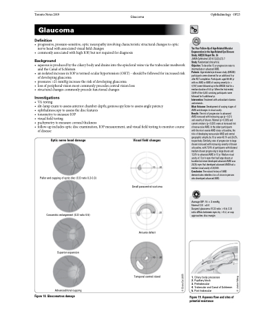
Average IOP: 15 ± 3 mmHg
Normal C:D : ≤0.4
Suspect glaucoma if C:D ratio >0.6, C:D ratio differs between eyes by >0.2, or cup approaches disc margin
543 2 1
1. Ciliary body processes
2. Pupillary block
3. Pretrabecular
4. Trabecular and Canal of Schlemm 5. Post-trabecular
Figure 19. Aqueous flow and sites of potential resistance
of disease
Optic nerve head damage
Pallor and cupping of optic disc (C:D ratio 0.2-0.3)
Concentric enlargement (C:D ratio 0.5)
Superior expansion
Advanced/total cupping
Figure 18. Glaucomatous damage
Visual field changes
10 20
Small paracentral scotoma
10 20
Arcuate defect
10 20
Temporal central island
© Diana Dai 2005
© Janice Wong
