
Page 942 - TNFlipTest
P. 942
OR8 Orthopedics
Orthopedic X-Ray Imaging
Toronto Notes 2019
CRPS/RSD
An exaggerated response to an insult in the extremities; characterized by symptoms
of hyperalgesia and allodynia, with signs
of autonomic dysfunction (temperature asymmetry, mottling, hair or nail changes)
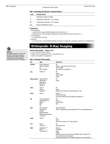
Table 4. Outerbridge Classification of Chondral Defects
Grade Chondral Damage
I Softening and swelling of cartilage
II Fragmentation and fissuring <1/2” in diameter
III Fragmentation and fissuring >1/2” in diameter
IV Erosion of cartilage down to bone
Treatment
• individualized
■ patient factors (age, skeletal maturity, activity level, etc.)
■ defect factors (Outerbridge Classification, subchondral bone involvement, etc.)
• non-operative
■ rest, NSAIDs, bracing
• operative
■ microfracture, osteochondral grafting (autograft or allograft), autologous chondrocyte implantation
Orthopedic X-Ray Imaging
General Principles - “Rule of 2s”
• x-ray1jointaboveand1below
• obtainatleast2orthogonalviews±specializedviews • 2sides,asneededforcomparison
Table 5. Orthopedic X-Ray Imaging
Site
Shoulder
Arm
Elbow/Forearm
Wrist Pelvis Hip
Knee
Ankle
Foot Spine
Injury
Anterior dislocation Posterior dislocation AC
Frozen shoulder
Humerus #
Supracondylar # Radial head # Monteggia # Night stick # Galeazzi #
Colles’ # Smith # Scaphoid #
Pelvic #
Femoral head/neck # Intertrochanteric # Arthritis
SCFE
FAI
Knee dislocation Femur/tibia #
Patella #
Patella dislocation Patella femoral syndrome Tibia shaft #
Ankle #
Talar # Calcanial #
Compression # Burst # Cervical spine #
X-Ray Views
AP
Axillary ± stress view with 10 lb in hand Trans-scapular
Zanca view (10-15 cephalic tilt)
AP
Lateral Trans-scapular Axillary
AP Lateral
AP
Lateral
Scaphoid (wrist extension and ulnar deviation x 2 wk)
AP pelvis
Inlet and outlet views
Judet views (obturator and iliac oblique for acetabular #)
AP
Lateral Frog-leg lateral Dunn
AP standing, lateral
Skyline – tangential view with knees flexed at 45° to see patellofemoral joint
AP
Lateral
Mortise view: ankle at 15° of internal rotation
AP
Lateral Harris axial
AP spine
AP odontoid
Lateral
Oblique
Swimmer’s view: lateral view with arm abducted 180° to evaluate C7-T1 junction if lateral view is inadequate
Lateral flexion/extension view: evaluate subluxation of cervical vertebrae
