
Page 941 - TNFlipTest
P. 941
Toronto Notes 2019 Articular Cartilage
6. Follow Up
■ evaluate stages of bone healing
7. Rehabilitation
■ recommend rehabilitation when appropriate to regain function and avoid joint stiffn
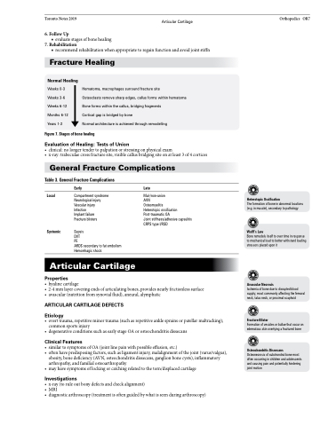
Fracture Healing
Orthopedics OR7
Normal Healing
Weeks 0-3 Weeks 3-6 Weeks 6-12 Months 6-12 Years 1-2
Hematoma, macrophages surround fracture site
Osteoclasts remove sharp edges, callus forms within hematoma Bone forms within the callus, bridging fragments
Cortical gap is bridged by bone
Normal architecture is achieved through remodelling
Figure 7. Stages of bone healing
Evaluation of Healing: Tests of Union
• clinical:nolongertendertopalpationorstressingonphysicalexam
• x-ray:trabeculaecrossfracturesite,visiblecallusbridgingsiteonatleast3of4cortices
General Fracture Complications
Table 3. General Fracture Complications
Local
Systemic
Early
Compartment syndrome Neurological injury Vascular injury
Infection
Implant failure Fracture blisters
Sepsis
DVT
PE
ARDS secondary to fat embolism Hemorrhagic shock
Late
Mal-/non-union
AVN
Osteomyelitis
Heterotopic ossification Post-traumatic OA
Joint stiffness/adhesive capsulitis CRPS type I/RSD
Heterotopic Ossification
The formation of bone in abnormal locations (e.g. in muscle), secondary to pathology
Wolff’s Law
Bone remodels itself to over time in response to mechanical load to better withstand loading stressors placed upon it
Avascular Necrosis
Ischemia of bone due to disrupted blood supply; most commonly affecting the femoral neck, talus neck, or proximal scaphoid
Fracture Blister
Formation of vesicles or bullae that occur on edematous skin overlying a fractured bone
Osteochondritis Dissecans
Osteonecrosis of subchondral bone most often occurring in children and adolescents and causing pain and potentially hindering joint motion
Articular Cartilage
Properties
• hyalinecartilage
• 2-4 mm layer covering ends of articulating bones, provides nearly frictionless surface • avascular(nutritionfromsynovialfluid),aneural,alymphatic
ARTICULAR CARTILAGE DEFECTS
Etiology
• overttrauma,repetitiveminortrauma(suchasrepetitiveanklesprainsorpatellarmaltracking); common sports injury
• degenerativeconditionssuchasearlystageOAorosteochondritisdissecans
Clinical Features
• similartosymptomsofOA(jointlinepainwithpossibleeffusion,etc.)
• oftenhavepredisposingfactors,suchasligamentinjury,malalignmentofthejoint(varus/valgus),
obesity, bone deficiency (AVN, osteochondritis dissecans, ganglion bone cysts), inflammatory
arthropathy, and familial osteoarthropathy
• may have symptoms of locking or catching related to the torn/displaced cartilage
Investigations
• x-ray(toruleoutbonydefectsandcheckalignment)
• MRI
• diagnosticarthroscopy(treatmentisoftenguidedbywhatisseenduringarthroscopy)
