Page 945 - TNFlipTest
P. 945
 Toronto Notes 2019 Shoulder
Clinical Features
• symptoms:painandfever
• onexam:erythema,tenderness,edemacommon±abscess/drainingsinustract;impairedfunction/WB
Diagnosis
• seeMedicalImaging,MI23
• workupincludes:WBCanddifferential,ESR,CRP,bloodculture,aspirateculture/bonebiopsy
Orthopedics OR11
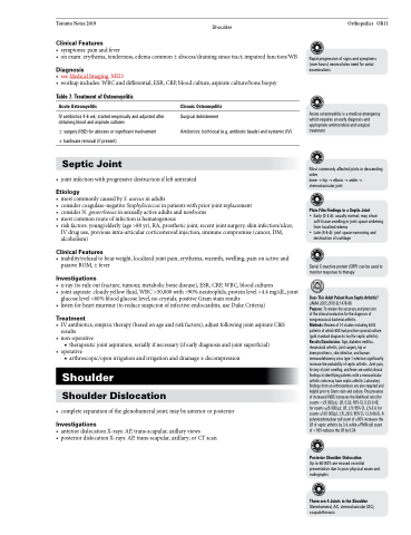
   Table 7. Treatment of Osteomyelitis
Acute Osteomyelitis
IV antibiotics 4-6 wk; started empirically and adjusted after obtaining blood and aspirate cultures
± surgery (I&D) for abscess or significant involvement ± hardware removal (if present)
Septic Joint
Chronic Osteomyelitis
Surgical debridement
Antibiotics: both local (e.g. antibiotic beads) and systemic (IV)
Rapid progression of signs and symptoms (over hours) necessitates need for serial examinations
Acute osteomyelitis is a medical emergency which requires an early diagnosis and appropriate antimicrobial and surgical treatment
Most commonly affected joints in descending order
knee→hip→elbow→ankle→ sternoclavicular joint
Plain Film Findings in a Septic Joint
• Early (0-3 d): usually normal; may show soft-tissue swelling or joint space widening from localized edema
• Late (4-6 d): joint space narrowing and destruction of cartilage
Serial C-reactive protein (CRP) can be used to monitor response to therapy
Does This Adult Patient Have Septic Arthritis?
JAMA 2007;297(13):1478-88
Purpose: To review the accuracy and precision
of the clinical evaluation for the diagnosis of nongonococcal bacterial arthritis.
Methods: Review of 14 studies including 6242 patients of which 653 had positive synovial culture (gold standard diagnostic tool for septic arthritis). Results/Conclusions: Age, diabetes mellitus, rheumatoid arthritis, joint surgery, hip or
knee prosthesis, skin infection, and human immunodeficiency virus type 1 infection significantly increase the probability of septic arthritis. Joint pain, history of joint swelling, and fever are useful clinical findings in identifying patients with a monoarticular arthritis who may have septic arthritis. Laboratory findings from an arthrocentesis are also required and helpful prior to Gram stain and culture. The presence of increased WBC increases the likelihood ratio (for counts <25 000/μL: LR, 0.32; 95% CI, 0.23-0.43; for counts ≥25 000/μL: LR, 2.9; 95% CI, 2.5-3.4; for counts ≥100 000/μL: LR, 28.0; 95% CI, 12.0-66.0). A polymorphonuclear cell count of ≥90% increases the LR of septic arthritis by 3.4, while a PMN cell count of <90% reduces the LR by 0.34.
Posterior Shoulder Dislocation
Up to 60-80% are missed on initial presentation due to poor physical exam and radiographs
There are 4 Joints in the Shoulder
Glenohumeral, AC, sternoclavicular (SC), scapulothoracic
           • jointinfectionwithprogressivedestructionifleftuntreated
Etiology
• mostcommonlycausedbyS.aureusinadults
• considercoagulase-negativeStaphylococcusinpatientswithpriorjointreplacement
• considerN.gonorrhoeaeinsexuallyactiveadultsandnewborns
• mostcommonrouteofinfectionishematogenous
• riskfactors:young/elderly(age>80yr),RA,prostheticjoint,recentjointsurgery,skininfection/ulcer,
IV drug use, previous intra-articular corticosteroid injection, immune compromise (cancer, DM, alcoholism)
Clinical Features
• inability/refusaltobearweight,localizedjointpain,erythema,warmth,swelling,painonactiveand passive ROM, ± fever
Investigations
• x-ray(toruleoutfracture,tumour,metabolicbonedisease),ESR,CRP,WBC,bloodcultures
• jointaspirate:cloudyyellowfluid,WBC>50,000with>90%neutrophils,proteinlevel>4.4mg/dL,joint
glucose level <60% blood glucose level, no crystals, positive Gram stain results
• listenforheartmurmur(toreducesuspicionofinfectiveendocarditis,useDukeCriteria)
Treatment
• IVantibiotics,empirictherapy(basedonageandriskfactors),adjustfollowingjointaspirateC&S results
• non-operative
■ therapeutic joint aspiration, serially if necessary (if early diagnosis and joint superficial)
• operative
■ arthroscopic/open irrigation and irrigation and drainage ± decompression
Shoulder
Shoulder Dislocation
• completeseparationoftheglenohumeraljoint;maybeanteriororposterior
Investigations
• anteriordislocationX-rays:AP,trans-scapular,axillaryviews
• posteriordislocationX-rays:AP,trans-scapular,axillary;orCTscan


