
Page 946 - TNFlipTest
P. 946
OR12 Orthopedics
Shoulder
Toronto Notes 2019
Table 8. Anterior and Posterior Shoulder Dislocation
Shoulder passive ROM: abduction – 180°, adduction – 45°, flexion – 180°, extension – 45°, int. rotation – level of T4, ext. rotation – 40-45°
Factors Causing Shoulder Instability
• Shallow glenoid
• Loose capsule
• Ligamentous laxity
Frequency of Dislocations
• Anterior shoulder > Posterior shoulder
• Posterior hip > Anterior hip
The glenohumeral joint is the most commonly dislocated joint in the body since stability is sacrificed for motion
MECHANISM
Anterior Shoulder Dislocation (>90%)
Abducted arm is externally rotated/hyperextended Blow to posterior shoulder
Involuntary, usually traumatic; voluntary, atraumatic
Posterior Shoulder Dislocation (5%)
Adducted, internally rotated, flexed arm FOOSH
3 Es (epileptic seizure, EtOH, electrocution) Blow to anterior shoulder
Pain, arm is held in adduction and internal rotation; external rotation is blocked
Anterior shoulder flattening, prominent coracoid, palpable mass posterior to shoulder
Positive posterior apprehension (“jerk”) test: with patient supine, flex elbow 90° and adduct, internally rotate the arm while applying a posterior force to the shoulder; patient will “jerk” back with the sensation of subluxation
Note: the posterior apprehension test is used to test for recurrent posterior instability, NOT for acute injury
Full neurovascular exam as per anterior shoulder dislocation
Humeral head is posterior
Humeral head is posterior to centre of “Mercedes-Benz” sign
Partial vacancy of glenoid fossa (vacant glenoid sign) and >6 mm space between anterior glenoid rim and humeral head (positive rim sign), humeral head may resemble a lightbulb due to internal rotation (lightbulb sign)
± Reverse Hill-Sachs lesion (75% of cases): divot in anterior humeral head
± Reverse bony Bankart lesion: avulsion of the posterior glenoid labrum from the bony glenoid rim
Closed reduction with sedation and muscle relaxation Inferior traction on a flexed elbow with pressure on the back of the humeral head
Obtain post-reduction x-rays
Check post-reduction NVS
Sling in abduction and external rotation x 3 wk, followed by shoulder rehabilitation (dynamic stabilizer strengthening)
CLINICAL FEATURES
5 6
4 3 2
Symptoms Shoulder Exam
Neurovascular Exam Including
Pain, arm slightly abducted and externally rotated with inability to internally rotate
“Squared off” shoulder
Positive apprehension test: patient looks apprehensive with gentle shoulder abduction and external rotation to 90o as humeral head is pushed anteriorly and recreates feeling of anterior dislocation Positive relocation test: a posteriorly directed
force applied during the apprehension test relieves apprehension since anterior subluxation is prevented Positive sulcus sign: presence of subacromial indentation with distal traction on humerus indicates inferior shoulder instability (
Axillary nerve: sensory patch over deltoid and deltoid contraction
Musculocutaneous nerve: sensory patch on lateral forearm and biceps contraction
1
789
Axillary View
Trans-scapular ‘Y’ View
AP View
Hill-Sachs and Bony Bankart Lesions
TREATMENT
Humeral head is anterior
Humeral head is anterior to the centre of the “Mercedes-Benz”sign
Sub-coracoid lie of the humeral head is most common
± Hill-Sachs lesion: compression fracture of posterior humeral head due to forceful impaction of an anteriorly dislocated humeral head against the glenoid rim
± bony Bankart lesion: avulsion of the anterior glenoid labrum (with attached bone fragments) from the glenoid rim
Closed reduction with IV sedation and muscle relaxation
Traction-countertraction: assistant stabilizes torso with a folded sheet wrapped across the chest while the surgeon applies gentle steady traction
Stimson: while patient lies prone with arm hanging over table edge, hang a 5 lb weight on wrist for 15-20 min
Hippocratic method: place heel into patient’s axilla and apply traction to arm
Cunningham’s method: low risk, low pain; if not successful try above methods
Obtain post-reduction x-rays
Check post-reduction NVS
Sling x 3 wk (avoid abduction and external rotation), followed by shoulder rehabilitation (dynamic stabilizer strengthening)
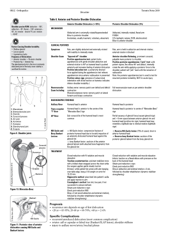
1. Manubrium
2. Sternoclavicular joint 3. Clavicle
4. Coracoid process
5. AC joint
6. Acromion
7. Humerus
8. Glenohumeral joint 9. Scapula
Figure 9. Shoulder joints
Figure 10. Mercedes-Benz
Bankart
Hill-Sachs
Figure 11. Posterior view of anterior dislocation causing Hill-Sachs and Bankart lesions
Prognosis
RADIOGRAPHIC FINDINGS
Coracoid process
Acromion
Scapula Humerus
• recurrenceratedependsonageoffirstdislocation
• <20yr=65-95%;20-40yr=60-70%;>40yr=2-4%
Specific Complications
• recurrent/unreduceddislocation(mostcommoncomplication)
• rotatorcufforcapsularorlabraltear(Bankart/SLAPlesion),shoulderstiffness • injurytoaxillarynerve/artery,brachialplexus
© Mary Sims 2003 © Kajeandra Ravichandiran 2012
© Jason Raine
