Page 1290 - TNFlipTest
P. 1290
 RH4 Rheumatology
Synovial Fluid Analysis
Toronto Notes 2019
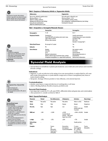
Table 6. Symptoms of Inflammatory Arthritis vs. Degenerative Arthritis
   The presence of synovitis often indicates articular as opposed to non-articular joint pain; synovitis presents with: soft tissue swelling, effusion, warmth, and pain with movement
Inflammatory
Pain at rest, relieved by gentle motion Morning stiffness >1 h
Warmth, swelling, erythema Malalignment/deformity (late finding) Extra-articular manifestations Nighttime awakening due to pain
Degenerative
Pain with motion, relieved by rest
Morning stiffness <1/2 h
Joint instability, buckling, locking
Bony enlargement, malalignment/deformity (late finding) Evening/end of day pain
  Table 7. Seropositive vs. Seronegative Rheumatic Diseases
    CRP
Advantages
• Unaffected by age/gender
• Rapid response to inflammatory stimulus • Wide range of clinically relevant values • Can be measured on stored sera
• Quantification precise/reproducible Disadvantages
• More expensive
Demographics Peripheral Arthritis
Pelvic/Axial Disease Enthesitis Extra-Articular
Seropositive
F>M
Symmetrical
Small (PIP, MCP) and medium joints (wrist, knee, ankle, elbow) common
DIP less often involved
No (except for C-spine)
No
Nodules
Vasculitis
Sicca
Raynaud’s phenomenon
Rashes, internal organ involvement (lung, cardiac) Entrapment neuropathies (i.e. carpal tunnel syndrome)
Seronegative
M>F
Usually asymmetrical
Usually larger joints, lower extremities (exception: PsA)
DIP in PsA
Dactylitis (“sausage digit”)
Yes Yes
Iritis (anterior uveitis)
Oral ulcers
Gastrointestinal Dermatological (psoriasis or keratodermia) Genitourinary inflammation
  Synovial Fluid Analysis
           Most Important Tests of Synovial Fluid (3 Cs)
1. Culture and gram stain
2. Cell count and differential
3. Crystal examination (protein, LDH, glucose less helpful)
Choosing Wisely Canada Recommendations
1. Do not order ANA as a screening test
in patients without specific signs or
symptoms of SLE or another CTD 2. Do not order an HLA-B27 unless
spondyloarthritis is suspected based on
specific signs or symptoms
3. Do not repeat DEXA scans more often than
every 2 years
4. Do not prescribe bisphosphonates for
patients at low risk of fracture
5. Do not perform whole body bone scans
(e.g. scintigraphy) for diagnostic screening for peripheral and axial arthritis in the adult population
• synovialfluidisanultrafiltrateofplasmaplushyaluronicacid;itlubricatesjointsurfacesandnourishes articular cartilage
Indications
• diagnostic:toruleoutinfectioninthesettingofanacutemonoarthritis;toanalyzefluidforcellcount and crystals if hemarthrosis or crystal arthritis is suspected or if there is unexplained joint, bursa or tendon sheath swelling
• therapeutic:drainageofblood,purulentortenseeffusions;corticosteroidinjection
Contraindications
• absolute:openlesionorsuspectedinfectionofoverlyingskinorsofttissue • relative:bleedingdiathesis,thrombocytopenia,prostheticjoint
Synovial Fluid Analysis
• mostimportanttoassessthe3C’s:cellcount(WBC),differentialcultureandgramstain,andcrystalanalysis • otherparameterstoconsiderarelistedinTable8
Table 8. Synovial Fluid Analysis
 Parameter
Colour Clarity Viscosity
WBC/mm3
% PMN
Culture/Gram Stain
Examples
Normal
Pale yellow Clear
High
<200
<25% –
Non-Inflammatory
Pale yellow Clear
High
<2,000
<25% –
Trauma
OA Neuropathy Hypertrophic – arthropathy
Inflammatory
Pale yellow Opaque Low
≥2,000
(crystal induced arthritis – often much higher than 2,000)
≥25% –
Seropositive Seronegative Crystal arthropathies
Infectious
Yellow to white Opaque
Low or paradoxically high if purulent
>2000 (>50,000)
>75%
Usually positive
S. aureus
Gram negative GC → difficult
to culture, (may have a low WBC)
Hemorrhagic
Red/brown Sanguinous Variable
Variable
Variable –
Trauma Hemophilia


