
Page 1339 - TNFlipTest
P. 1339
Toronto Notes 2019
Urological Neoplasms
Urology U21
Urological Neoplasms
Approach to Renal Mass
Cystic
Solid
CT (exclude angiomyolipoma)
There is controversy over optimal management of small renal masses
Percutaneous needle biopsies of cystic renal masses may lead to peritoneal seeding
Tuberous Sclerosis
• Syndrome characterized by mental retardation, epilepsy, and adenoma sebaceum
• 45-80%ofpatientsalsopresentwith angiomyolipomas which are often multiple and bilateral
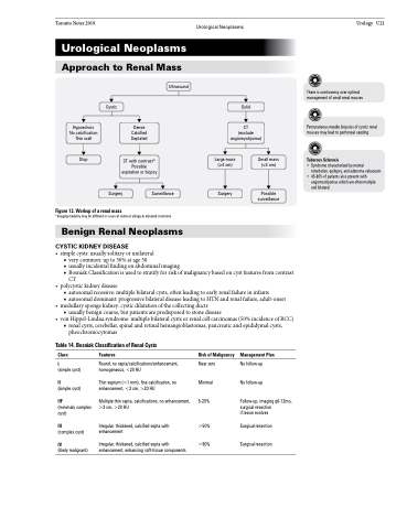
Ultrasound
Hypoechoic No calcification Thin wall
Stop
Dense Calcified Septated
CT with contrast* Possible aspiration or biopsy
Large mass (≥4 cm)
Surgery
Small mass (≤4 cm)
Possible surveillance
Figure 13. Workup of a renal mass
Surgery
Surveillance
*Imaging modality may be different in cases of contrast allergy or elevated creatinine
Benign Renal Neoplasms
CYSTIC KIDNEY DISEASE
• simplecysts:usuallysolitaryorunilateral
■ very common: up to 50% at age 50
■ usually incidental finding on abdominal imaging
■ Bosniak Classification is used to stratify for risk of malignancy based on cyst features from contrast
CT
• polycystickidneydisease
■ autosomal recessive: multiple bilateral cysts, often leading to early renal failure in infants
■ autosomal dominant: progressive bilateral disease leading to HTN and renal failure, adult-onset • medullaryspongekidney:cysticdilatationofthecollectingducts
■ usually benign course, but patients are predisposed to stone disease
• vonHippel-Lindausyndrome:multiplebilateralcystsorrenalcellcarcinomas(50%incidenceofRCC)
■ renal cysts, cerebellar, spinal and retinal hemangioblastomas, pancreatic and epididymal cysts, pheochromocytomas
Table 14. Bosniak Classification of Renal Cysts
Class
I
(simple cyst)
II
(simple cyst)
IIF
(minimaly complex cyst)
III
(complex cyst)
IV
(likely malignant)
Features
Round, no septa/calcifications/enhancement, homogeneous, <20 HU
Thin septum (<1 mm), fine calcification, no enhancement, <3 cm, >20 HU
Multiple thin septa, calcifications, no enhancement, >3 cm, >20 HU
Irregular, thickened, calcified septa with enhancement
Irregular, thickened, calcified septa with enhancement, enhancing soft-tissue components
Risk of Malignancy
Near zero Minimal 5-20%
>50% >90%
Management Plan
No follow-up No follow-up
Follow-up, imaging q6-12mo, surgical resection
if lesion evolves
Surgical resection Surgical resection
