
Page 1340 - TNFlipTest
P. 1340
U22 Urology
Urological Neoplasms
Toronto Notes 2019
Gerota’s fascia
T4
Table 15. Benign Renal Masses
T3 T2
T1
≤7 cm
Adrenal gland
Vein
Artery Ureter
© Carly Vanderlee
Epidemiology
Characteristics
Diagnosis
Management
Angiomyolipoma (Renal Hamartoma)
<1% of adult renal tumours
F>M
20% associated with tuberous sclerosis (especially if multiple, recurrent)
Clonal neoplasm consisting of blood vessels (angio-), smooth muscle (-myo-), and fat (-lipoma)
May extend into regional lymphatics and other organs and become symptomatic
Incidental finding on CT
Negative attenuation (-20 HU) on CT is pathognomonic Rare presentation of hematuria, flank pain, and palpable mass (same as RCC)
May consider surgical excision or embolization if symptomatic (pain, bleeding) or higher risk of bleeding (e.g. pregnancy)
Potential role for mTOR inhibitors in unresectable/ metastatic disease
Follow with serial U/S
Renal Oncocytoma
3-7% of renal tumours
M>F
Oncocytomas also found in adrenal, thyroid and parathyroid glands
Spherical, capsulated with possible central scar Histologically organized aggregates of eosinophilic cells originating from intercalated cells of collecting duct
Incidental finding on CT
Difficult to distinguish from RCC on imaging – treated as RCC until proven otherwise
Biopsy may be performed to rule out malignancy
Partial/radical nephrectomy for large masses HIFU or RFA for smaller masses
>7 cm
Renal cortex
Renal medulla
Renal capsule
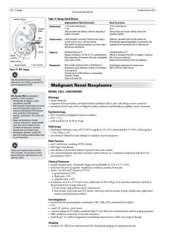
Figure 14. RCC staging
Role of environmental exposures (aromatic hydrocarbons, etc.) remains an unproven risk factor for development of RCC
RCC Systemic Effects: paraneoplastic syndromes (10-40% of patients)
• Hematopoietic disturbances: anemia,
polycythemia, raised ESR
• Endocrinopathies: hypercalcemia (increased
vitamin D hydroxylation), erythrocytosis (increased erythropoietin), HTN (increased renin), production of other hormones (prolactin, gonadotropins, TSH, insulin, and cortisol)
• Hepatic cell dysfunction or Stauffer syndrome: abnormal LFTs, decreased WBC count, fever, areas of hepatic necrosis;
no evidence of metastases; reversible following removal of primary tumour
• Hemodynamic alterations: systolic HTN (due to AV shunting), peripheral edema (due to caval obstruction)
Malignant Renal Neoplasms
RENAL CELL CARCINOMA
Etiology
• causeunknown
• originatesfromproximalconvolutedtubuleepithelialcellsinclearcellsubtype(mostcommon)
• hereditaryformsseenwithvonHippel-Lindausyndromeandhereditarypapillaryrenalcarcinoma
Epidemiology
• 85%ofprimarymalignanttumoursinkidney • M:F=1.5:1
• peakincidenceat50-60yrofage
Pathology
• histologicalsubtypes:clearcell(75-85%),papillary(10-15%),chromophobic(5-10%),collectingduct (<1%), other (<1%)
• sarcomatoidelementsinanysubtypeisamarkerofpoorprognosis
Risk Factors
• top3riskfactors:smoking,HTN,obesity
• end-stagerenaldisease
• miscellaneous: horseshoe kidney, acquired renal cystic disease
• role of environmental exposures (aromatic hydrocarbons, etc.) remains an unproven risk factor for
development of RCC
Clinical Features
• usuallyasymptomatic:frequentlydiagnosedincidentallybyU/SorCT(>50%) • indicatorsforpoorprognosis:weightloss,weakness,anemia,bonepain
• classic“toolatetriad”foundin10-15%
■ gross hematuria 50% ■ flank pain <50%
■ palpable mass <30%
• metastases:seenina1/3ofnewcases;additional20-40%willgoontodevelopmetastases(mostlyin late presentations or large tumours)
■ bone, brain, lung and liver most common site
■ may invade renal veins and IVC lumen. This may result in ascites, hepatic dysfunction, right atrial
tumour, and pulmonary emboli
Investigations
• routinelabsforparaneoplasticsyndromes(CBC,ESR,LFTs,extendedelectrolytes)
• U/A
• renalU/S:solidvs.cysticlesion
• contrast-enhancedCT:highersensitivitythanU/Sfordetectionofrenalmassesandforstagingpurposes • MRI:usefulforevaluationofvascularextension
• renalbiopsy:toconfirmdiagnosisifconsideringobservationorothernon-surgicaltherapy
Staging
• involvesCT,CXR,liverenzymesandLFTs,bone/headimaging(ifsymptomsdictate)
Tumour may invade renal veins and inferior vena cava lumen. This may result in ascites, hepatic dysfunction, right atrial tumour, and pulmonary emboli
