
Page 1341 - TNFlipTest
P. 1341
Toronto Notes 2019 Urological Neoplasms
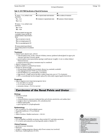
Table 16. 2010 TNM Classification of Renal Cell Carcinoma
TNM
Urology U23
T1: tumour <7 cm, confined to renal parenchyma
T1a: <4 cm T1b: 4-7 cm
T2: tumour >7 cm, confined to renal parenchyma
T2a: 7-10 cm T2b: >10cm
T3: tumour extends into major veins or perinephric tissues, but NOT into ipsilateral adrenal or beyond Gerota’s fascia
T3a: into renal vein or sinus fat T3b: into infradiaphragmatic IVC T3c: into supradiaphragmatic IVC
T4: tumour extends beyond Gerota’s fascia including extension into ipsilateral adrenal
Treatment
N0: no regional lymph node metastasis N1: metastasis in regional lymph nodes
M0: no evidence of metastasis M1: presence of distant metastasis
• surgical (open, laparoscopic, robotic)
■ radical nephrectomy: en bloc removal of kidney, tumour, ipsilateral adrenal gland (in upper pole
tumours) and intact Gerota’s capsule
■ partial nephrectomy (parenchyma-sparing): small tumour (roughly <4 cm) or solitary kidney/
bilateral tumours
■ surgical removal of solitary metastasis may be considered
• ablativetechniques(RFA)
• palliativeradiationtopainfulbonylesions • therapyforadvancedstage
■ tyrosine kinase inhibitors for metastatic disease (e.g. sunitinib, sorafenib)
■ anti-angiogenesis/anti-VEGF (e.g. bevacizumab)
■ mTOR inhibitors (e.g. temsirolimus, everolimus)
■ high-dose IL-2 (high toxicity but able to induce long-term cure in 5-7% of patients)
■ IFN-α: monotherapy has been largely replaced by molecularly targeted agents listed above
Prognosis
• stageatdiagnosismostimportantprognosticfactor ■ T1: 90-100% 5 yr survival
■ T2-T3: 60% 5 yr survival
■ metastatic disease: <5% 10 yr survival
Carcinoma of the Renal Pelvis and Ureter
Etiology
• riskfactorsinclude: ■ smoking
■ chemicals/dietary exposures (industrial dyes and solvents; aristolochic acid, aniline dyes) ■ analgesic abuse (acetaminophen, ASA, and phenacetin)
■ Balkannephropathy
■ prior exposure to cyclophosphamide
Epidemiology
• rare:accountsfor5%ofallurothelialcancers
• frequentlymultifocal,2-5%arebilateral
• M:F=3:1
• relativeincidence:bladder:renal:ureter=100:10:1
Pathology
• 85%arepapillaryurothelialcarcinoma;othersincludeSCCandadenocarcinoma • UCofureterandrenalpelvisarehistologicallysimilartobladderUC
Clinical Features
• gross/microscopichematuria
• flankpain
• storageorvoidingsymptoms(dysuriaonlyiflowerurinarytractinvolved) • flankmass±hydronephrosis(10-20%)
Differential Diagnosis of Filling Defect
• Urothelial carcinoma (differentiate via cytology and CT scan)
• Uric acid stone (differentiate via cytology and CT scan)
• Blood clot
• Pyelitis cystica
• Papillary necrosis
• Fungus ball
• Gas bubble from gas producing organisms
